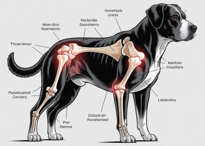
Understanding canine anatomy is crucial for anyone involved in animal care, from veterinarians to dedicated owners. Veterinary schools emphasize the importance of precise anatomical terminology. A key element within canine biomechanics concerns articulation; what is the shoulder joint of a canine called and how does it function? That answer relates directly to the scapula of the animal and the biomechanics behind that region of the anatomy.

The well-being of our canine companions relies heavily on our understanding of their physical structure. Canine anatomy, while complex, provides the foundation for recognizing potential health issues and ensuring a high quality of life for our dogs. Among the various anatomical components, the shoulder joint stands out as a critical area, essential for mobility and overall function.
But what exactly is the proper, formal name for this pivotal joint that allows our dogs to run, jump, and play?
The shoulder joint in canines is formally known as the Glenohumeral Joint. Understanding this term, and the anatomy it represents, is the first step toward appreciating the complexities and vulnerabilities of the canine shoulder.
To truly understand the Glenohumeral Joint, we need to lay the groundwork with a clear understanding of canine anatomy and the critical function this joint serves. Let’s dissect the anatomical foundations that make this essential joint so vital to our canine friends.
Decoding the Canine Shoulder: Anatomical Foundations
Understanding the shoulder joint requires a firm grasp of basic canine anatomy. Anatomy, the study of the body’s structure, gives us the language and the framework to understand how the different parts of a dog’s body work together. By appreciating the anatomy of the shoulder, we can better understand its normal function, recognize potential problems, and provide informed care.
Defining the Canine Shoulder Joint
The shoulder joint, or Glenohumeral Joint, is the point where the forelimb connects to the body. Unlike humans, dogs do not have a collarbone (clavicle) that directly connects the shoulder to the axial skeleton. Instead, the shoulder joint is primarily held in place by muscles, tendons, and ligaments, allowing for a great range of motion.
The primary functions of the shoulder joint are:
-
Mobility: Allowing for a wide range of movement, essential for running, jumping, and turning.
-
Weight-Bearing: Supporting the dog’s weight and absorbing impact during physical activity.
The Bones of the Canine Shoulder Joint
Two primary bones form the Glenohumeral Joint: the scapula and the humerus. Their unique shapes and interaction define the joint’s structure and determine its movement capabilities.
Scapula (Shoulder Blade)
The scapula, or shoulder blade, is a flat, triangular bone that lies on the upper part of the chest wall. It does not directly articulate with the spine, which contributes to the dog’s flexibility and range of motion in the forelimb.
A critical feature of the scapula is the glenoid fossa. This shallow, concave depression forms the "socket" portion of the shoulder joint. The glenoid fossa articulates with the head of the humerus.
Humerus (Upper Arm Bone)
The humerus is the long bone of the upper forelimb, extending from the shoulder to the elbow. The humeral head is the rounded, proximal end of the humerus, which fits into the glenoid fossa of the scapula.
This articulation between the humeral head and the glenoid fossa forms the Glenohumeral Joint, allowing for movement in multiple planes. The relatively shallow nature of the glenoid fossa, however, means that the joint relies heavily on surrounding soft tissues for stability.
Decoding the shoulder joint’s anatomical foundations lays the necessary groundwork, but to truly grasp its intricacies, we must now focus specifically on the Glenohumeral Joint itself. This is where the actual movement happens, where the bones meet, and where potential problems can arise.
The Glenohumeral Joint: A Closer Look
The term "Glenohumeral Joint" might sound complex, but it’s simply a descriptive name that tells us exactly what this joint connects: the glenoid fossa of the scapula and the humerus.
By understanding the etymology of this term, we gain a valuable insight into the anatomical structures involved.
Unpacking the Name: Gleno- and Humeral
Let’s break down the term "Glenohumeral" into its component parts. The prefix “gleno-” refers to the glenoid fossa, a shallow, concave depression on the scapula (shoulder blade). This fossa serves as the socket portion of the shoulder joint, accommodating the head of the humerus.
The term "humeral" quite simply relates to the humerus, which is the long bone of the upper arm. The rounded head of the humerus articulates with the glenoid fossa, forming the ball-and-socket structure of the Glenohumeral Joint.
Therefore, the name itself reveals the two primary bony components that constitute this crucial joint.
The Intricate Structure of the Glenohumeral Joint
The Glenohumeral Joint isn’t just about bone meeting bone; it’s a sophisticated structure designed for both mobility and stability. The interplay of cartilage, the joint capsule, and synovial fluid is essential for smooth, pain-free movement.
Cartilage: The Smooth Operator
Articular cartilage is a smooth, resilient tissue that covers the articulating surfaces of both the glenoid fossa and the humeral head. This cartilage reduces friction during movement, allowing the bones to glide effortlessly against each other.
Without healthy cartilage, bone-on-bone contact would occur, leading to pain, inflammation, and ultimately, degenerative joint disease.
The Joint Capsule: Encapsulating Stability
The entire Glenohumeral Joint is enclosed within a fibrous joint capsule. This capsule provides stability to the joint, preventing excessive movement and dislocation.
The capsule is composed of strong connective tissue that helps to hold the humerus in place within the glenoid fossa.
Synovial Fluid: The Lubricant of Motion
The joint capsule is lined with a synovial membrane, which produces synovial fluid. This viscous fluid acts as a lubricant, further reducing friction within the joint.
Synovial fluid also provides nutrients to the cartilage and removes waste products, contributing to the overall health of the joint.
In essence, the Glenohumeral Joint is a carefully engineered structure where bone, cartilage, the joint capsule, and synovial fluid work in perfect harmony. Any disruption to this delicate balance can lead to pain and impaired mobility, highlighting the importance of understanding its intricate anatomy.
The intricate structure of the Glenohumeral Joint, with its cartilage-lined surfaces and synovial fluid lubrication, provides a foundation for movement. However, this foundation requires a robust support system to function correctly. This support system, comprised of ligaments, tendons, and muscles, is what truly enables the shoulder joint to achieve its impressive range of motion while maintaining stability.
Support System: Ligaments, Tendons, and Muscles in Canine Shoulders
The canine shoulder joint’s impressive functionality stems not just from its bony architecture, but from the intricate interplay of ligaments, tendons, and muscles. These structures act as the joint’s support system, enabling a wide range of motion while ensuring stability. Understanding their individual roles and collective contribution is crucial for comprehending the overall biomechanics of the shoulder.
Ligaments: The Stabilizers
Ligaments are strong, fibrous bands of connective tissue that connect bone to bone. Their primary role is to provide stability to the joint by limiting excessive movement and preventing dislocations. In the canine shoulder, several key ligaments contribute to this stability.
Glenohumeral ligaments, for example, connect the humerus to the glenoid fossa, reinforcing the joint capsule and preventing excessive abduction (movement away from the body). Other important ligaments around the shoulder include the coracohumeral ligament, which connects the coracoid process of the scapula to the humerus, contributing to rotational stability. These ligaments work in concert to maintain the integrity of the joint during various activities.
Damage to these ligaments, often through trauma or repetitive strain, can lead to instability and pain, requiring veterinary intervention.
Tendons: The Force Transmitters
Tendons are tough, inelastic cords that connect muscles to bones. Unlike ligaments, which stabilize joints, tendons transmit the force generated by muscles to create movement. Several tendons play critical roles in the function of the canine shoulder.
The biceps brachii tendon, for instance, attaches the biceps muscle to the scapula, contributing to shoulder flexion (bending the joint) and stabilization. The supraspinatus tendon is another key structure, attaching the supraspinatus muscle to the humerus and assisting in shoulder abduction and stabilization.
Tendon injuries, such as strains or tears, are common in active dogs and can significantly impact their mobility.
Muscles: The Prime Movers
Muscles are the powerhouses behind shoulder movement. They contract to generate force, which is then transmitted through tendons to move the bones of the joint. Numerous muscles surround the canine shoulder, each contributing to specific movements and overall stability.
The deltoid muscle is a major abductor and flexor of the shoulder, allowing the dog to lift its leg to the side and forward. The supraspinatus and infraspinatus muscles, part of the rotator cuff, are crucial for external rotation and stabilization of the humeral head within the glenoid fossa. The subscapularis muscle, another rotator cuff muscle, provides internal rotation and further stabilizes the joint.
The coordinated action of these and other muscles enables the wide range of motion characteristic of the canine shoulder. Weakness or imbalance in these muscles can lead to instability and increase the risk of injury.
The Symphony of Movement: How Muscles, Tendons, and Ligaments Interact
The shoulder’s range of motion and stability are not solely dependent on any one structure, but rather on the coordinated action of all three. Ligaments provide passive stability, preventing excessive movement. Tendons act as the essential link, that allow muscles to provide controlled movement. Muscles generate the force and control the direction of movement, while tendons transmit that force to the bones.
For example, when a dog reaches forward, the deltoid and biceps brachii muscles contract, pulling on their respective tendons. This force causes the humerus to flex at the shoulder joint. Simultaneously, the glenohumeral ligaments tighten to prevent excessive forward movement, and the rotator cuff muscles contract to stabilize the humeral head within the glenoid fossa, preventing dislocation.
This intricate interplay ensures smooth, controlled movement while maintaining the integrity of the joint. Any disruption to this system, whether through injury to a ligament, tendon, or muscle, can compromise shoulder function and lead to pain and lameness. A comprehensive understanding of these supporting structures is essential for diagnosing and treating canine shoulder problems.
The intricate dance of ligaments, tendons, and muscles allows the canine shoulder its remarkable flexibility. However, this complex system is also vulnerable to a range of issues that can compromise its function.
Common Canine Shoulder Joint Issues
The canine shoulder joint, though a marvel of biomechanical engineering, is susceptible to various ailments. These problems can range from developmental abnormalities to degenerative conditions, significantly impacting a dog’s quality of life. Understanding these common issues is crucial for early detection, appropriate management, and ultimately, ensuring the well-being of our canine companions.
Shoulder Dysplasia: A Developmental Challenge
Shoulder dysplasia is a developmental condition characterized by an abnormality in the formation of the shoulder joint. Specifically, it often involves a shallow glenoid fossa, the socket that receives the head of the humerus.
This shallowness leads to instability within the joint. This instability can result in abnormal wear and tear, predisposing the dog to early-onset osteoarthritis and pain. Certain breeds, particularly larger breeds, are more prone to shoulder dysplasia due to their rapid growth rates and genetic predispositions.
Osteoarthritis: The Wear and Tear of Time (and Injury)
Osteoarthritis, also known as degenerative joint disease, is a progressive condition where the cartilage within the joint gradually breaks down. This breakdown leads to inflammation, pain, and reduced range of motion.
In the canine shoulder, osteoarthritis can arise from various factors, including:
-
Age-related degeneration: Natural wear and tear over time.
-
Previous injuries: Such as dislocations or fractures.
-
Underlying conditions: Like shoulder dysplasia, which accelerates cartilage damage.
The impact of osteoarthritis can be significant, limiting a dog’s ability to engage in normal activities and causing chronic discomfort. Management typically involves pain relief, weight management, and strategies to support joint health.
Ligament Injuries: Disrupting Stability
Ligaments play a crucial role in maintaining the stability of the shoulder joint. Injuries to these ligaments, such as sprains or tears, can lead to instability and pain.
One common ligament injury is a luxation, or dislocation, of the shoulder joint. This occurs when the humerus pops out of the glenoid fossa. Luxations can result from trauma, such as a fall or direct impact.
Other ligament injuries can develop gradually due to repetitive strain or overuse. Prompt veterinary attention is crucial for ligament injuries. This ensures appropriate diagnosis, stabilization, and rehabilitation to restore shoulder function.
Diagnosis and Treatment: The Veterinarian’s Role
The preceding discussion highlighted the common issues that can plague the canine shoulder joint. But what happens when you suspect your furry friend is suffering from one of these ailments? The answer lies in the expertise of a veterinarian. From initial assessment to implementing a tailored treatment plan, a veterinarian plays a pivotal role in restoring your dog’s comfort and mobility.
Identifying the Problem: The Veterinarian’s Approach
Veterinarians employ a multifaceted approach to identify shoulder problems in canines. They understand that accurate diagnosis is the cornerstone of effective treatment. This process typically begins with a thorough history and physical examination.
The veterinarian will ask detailed questions about your dog’s symptoms, including:
- When the lameness started
- What activities exacerbate the pain
- If there has been any previous trauma
This information helps narrow down the potential causes of the problem.
Diagnostic Tools: Unveiling the Underlying Cause
The physical examination is a crucial step, involving careful palpation of the shoulder joint.
The veterinarian will assess the range of motion, looking for any restrictions or pain upon manipulation.
They may also perform specific orthopedic tests to evaluate the stability of the ligaments and tendons surrounding the joint.
However, physical exams alone may not provide a complete picture.
Imaging techniques are often necessary to visualize the underlying structures and confirm the diagnosis.
-
X-rays (radiographs) are commonly used to assess bone structures and identify fractures, arthritis, or bone tumors.
-
More advanced imaging, such as MRI (magnetic resonance imaging), provides detailed images of soft tissues, including ligaments, tendons, and cartilage. MRI is particularly useful for diagnosing ligament tears or cartilage damage that may not be visible on X-rays.
Treatment Options: A Path to Recovery
Once a diagnosis has been established, the veterinarian will develop a treatment plan tailored to your dog’s specific condition. Treatment options vary depending on the severity and nature of the problem.
Conservative Management
In many cases, conservative management is the first line of defense.
This approach focuses on alleviating pain and inflammation, and promoting healing.
-
Medications play a crucial role, with pain relievers (analgesics) and anti-inflammatory drugs (NSAIDs) commonly prescribed to reduce discomfort and swelling.
-
Physical therapy can also be beneficial, helping to improve range of motion, strengthen surrounding muscles, and promote joint stability. This may involve exercises, massage, and hydrotherapy.
Surgical Intervention
When conservative treatments fail to provide adequate relief, or in cases of severe injury, surgery may be necessary. Several surgical options are available, depending on the specific problem.
-
Arthroscopy is a minimally invasive technique that allows the veterinarian to visualize and treat joint problems using a small camera and specialized instruments. This approach is often used to repair ligament tears or remove damaged cartilage.
-
In cases of severe osteoarthritis, joint replacement may be considered. This involves replacing the damaged shoulder joint with an artificial joint, restoring pain-free movement.
Long-Term Management and Monitoring
Regardless of the treatment approach, ongoing monitoring and management are essential for maintaining your dog’s shoulder health.
This may involve regular veterinary checkups, continued medication, and modifications to your dog’s activity level.
Weight management is also crucial, as excess weight can place additional stress on the shoulder joint.
By working closely with your veterinarian, you can help ensure that your canine companion enjoys a long and active life, free from the pain and limitations of shoulder joint problems.
FAQs: Canine Shoulder Anatomy Explained
Here are some frequently asked questions about the canine shoulder joint, also known as the scapulohumeral joint. We hope these answers provide clarity and help you understand your dog’s anatomy better.
What exactly is the shoulder joint in a dog?
The shoulder joint, or scapulohumeral joint, is the connection between the scapula (shoulder blade) and the humerus (upper arm bone) in a canine. It’s a ball-and-socket joint, allowing for a wide range of motion.
Why is knowing the name of the canine shoulder joint important?
Understanding that the shoulder joint of a canine is called the scapulohumeral joint allows for more effective communication with veterinarians and other pet professionals. Using correct terminology helps ensure accurate diagnoses and treatment plans.
What types of movements are enabled by the canine shoulder joint?
The scapulohumeral joint allows for flexion, extension, abduction (moving away from the body), adduction (moving towards the body), circumduction, and rotation. This wide range of motion is crucial for a dog’s locomotion and agility.
What are some common problems affecting the shoulder joint of a canine?
Common issues affecting the shoulder joint of a canine, or the scapulohumeral joint, include shoulder instability, osteoarthritis, and injuries like sprains and strains. Early diagnosis and treatment are essential for managing these conditions.
So, now you know more about what is the shoulder joint of a canine called! Hopefully, this has helped you better understand your furry friend’s body a little better.


