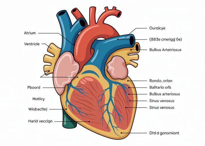
The single-loop circulatory system, a hallmark of fish anatomy, presents a fascinating subject of study often visualized using a fish heart diagram labeled. Understanding this circulatory system is crucial for anyone studying Ichthyology, the branch of zoology dedicated to fish. The structure of the fish heart itself, consisting of two chambers, is elegantly illustrated in any properly labeled diagram. Furthermore, the scientific community utilizes these diagrams to teach and research cardiovascular function across different species.

The heart, a tireless engine of life, beats within every creature, silently orchestrating the flow of existence. While we often marvel at the complexity of the human heart, the fish heart, seemingly simpler in design, holds its own secrets to survival in an aquatic world. Understanding this vital organ unveils a fascinating glimpse into the elegant adaptations that allow fish to thrive in their watery realms.
The Fish Heart: A Cornerstone of Aquatic Life
The fish heart plays a crucial role in maintaining life underwater. It’s the central pump responsible for circulating blood, delivering oxygen and nutrients, and removing waste products from tissues.
Without a properly functioning heart, a fish cannot sustain the energy demands of swimming, feeding, and reproducing. The fish heart, therefore, is fundamental to its survival.
The Fish Circulatory System: A Unique Design
The fish circulatory system boasts unique features optimized for aquatic life. It’s a single-loop system, meaning blood passes through the heart only once per circuit of the body. This differs significantly from the double-loop system found in mammals and birds.
Another key feature lies in the intimate relationship between the heart and the gills. The heart pumps blood directly to the gills. This is where gas exchange occurs, and the blood becomes oxygenated.
To truly appreciate the intricacies of this system, a labeled diagram serves as an invaluable tool. Visualizing the flow of blood through the heart’s chambers and the network of blood vessels provides a clearer understanding of the fish’s circulatory process.
A Journey into the Fish Heart: Structure, Function, and Significance
This guide embarks on a detailed exploration of the fish heart. We will delve into its anatomical structure, dissecting the roles of each component.
We will examine its functional mechanics, tracing the path of blood flow through its chambers.
Finally, we will consider its significance. It is of relevance to understanding fish physiology, ecological adaptations, and broader biological principles. Prepare to uncover the wonders of this remarkable organ and gain a deeper appreciation for the ingenuity of life beneath the waves.
The heart, a tireless engine of life, beats within every creature, silently orchestrating the flow of existence. While we often marvel at the complexity of the human heart, the fish heart, seemingly simpler in design, holds its own secrets to survival in an aquatic world. Understanding this vital organ unveils a fascinating glimpse into the elegant adaptations that allow fish to thrive in their watery realms.
The Basic Anatomy of a Fish Heart: A Two-Chambered Wonder
The fish heart, in its elegant simplicity, stands as a testament to evolutionary efficiency. While mammalian hearts boast four chambers, the fish heart operates effectively with just two: the atrium and the ventricle. These chambers, along with the sinus venosus and bulbus arteriosus, orchestrate the circulatory process, ensuring the delivery of oxygen and nutrients to the fish’s tissues. Understanding the function of each component is key to appreciating the heart’s overall role in sustaining aquatic life.
The Two Chambers: Atrium and Ventricle
The fish heart is often described as a two-chambered pump. This description, while accurate, can be misleading without understanding the roles of the additional components that contribute to its function.
The atrium serves as the receiving chamber for deoxygenated blood returning from the body.
It’s a thin-walled structure designed to efficiently collect blood before passing it on to the ventricle.
The ventricle, in contrast, is a thick-walled, muscular chamber responsible for pumping blood towards the gills, where oxygenation occurs. This powerful contraction is essential for driving blood through the circulatory system.
Key Components: Sinus Venosus and Bulbus Arteriosus
Beyond the two primary chambers, two other structures play crucial roles in the fish heart’s operation: the sinus venosus and the bulbus arteriosus.
The sinus venosus is a thin-walled sac that collects deoxygenated blood from the fish’s veins before it enters the atrium. It acts as a reservoir, ensuring a smooth and continuous flow of blood into the heart. This also helps with regulating blood volume and pressure.
The bulbus arteriosus, found in most fish (though replaced by the conus arteriosus in some), is an elastic chamber that receives blood from the ventricle and helps to smooth out the pulsatile flow before it enters the gills. Think of it as a hydraulic accumulator. This is particularly important for protecting the delicate gill capillaries from pressure surges.
Visualizing the Fish Heart: A Labeled Diagram
To fully grasp the spatial relationships and functions of these components, a labeled diagram of the fish heart is invaluable. The diagram should clearly illustrate the atrium, ventricle, sinus venosus, and bulbus arteriosus, highlighting their connections and relative positions. By visualizing the flow of blood through these structures, one can develop a deeper understanding of the fish heart’s remarkable design.
The elegant dance of contraction and relaxation within the fish heart sets the stage for an equally fascinating journey: the circulatory system. Unlike the complex, multi-looped systems found in mammals and birds, the fish circulatory system operates on a single, efficient loop. This design reflects the unique physiological demands of aquatic life, where oxygen uptake occurs directly from the water through the gills.
The Fish Circulatory System: A Single-Loop Journey
The fish circulatory system distinguishes itself through its single-loop design. This means that blood passes through the heart only once during each complete circuit of the body. Understanding this fundamental difference from the circulatory systems of terrestrial vertebrates is key to appreciating the efficiency and elegance of the fish’s adaptation to its aquatic environment.
The Single-Loop System: Heart to Gills to Body
In a single-loop system, blood follows a direct path: from the heart to the gills, then to the body, and finally back to the heart. The heart pumps deoxygenated blood to the gills.
At the gills, blood picks up oxygen and releases carbon dioxide. Then, oxygenated blood flows from the gills, it circulates through the body’s tissues, delivering oxygen and nutrients.
Finally, after delivering oxygen to the tissues, the now deoxygenated blood returns to the heart, completing the circuit.
The Gills: Orchestrating Gas Exchange
The gills are the linchpin of the fish circulatory system. Their primary function is gas exchange, which is the process of extracting oxygen from the water and releasing carbon dioxide into it.
This exchange occurs as water flows over the gill filaments, which are highly vascularized structures. The efficiency of this process is enhanced by the countercurrent exchange system, where blood flows in the opposite direction to water, maximizing oxygen uptake.
Blood Vessels within the Gills: Facilitating Exchange
Within the gills, a dense network of capillaries lines the gill filaments. These tiny blood vessels bring blood into close proximity with the water, facilitating the diffusion of gases.
Oxygen-rich water passes over the capillaries, and oxygen diffuses into the blood, while carbon dioxide moves out of the blood into the water. This exchange is vital for the fish’s survival, providing the necessary oxygen for cellular respiration.
Blood Vessels: Transporting Life’s Essence
Beyond the gills, blood vessels play a crucial role in transporting blood throughout the fish’s body. Arteries carry oxygenated blood away from the gills, branching into smaller arterioles that deliver blood to capillaries within tissues and organs.
Capillaries are the sites of nutrient and waste exchange, where oxygen and nutrients are delivered to cells, and carbon dioxide and metabolic waste products are picked up.
From the capillaries, blood flows into venules, which merge into larger veins that carry deoxygenated blood back to the heart, completing the circulatory loop. The intricate network of blood vessels ensures that every cell in the fish’s body receives the oxygen and nutrients it needs to function.
The intricate workings of the gills highlighted the vital role they play in oxygenating the blood, setting the stage for its journey throughout the fish’s body. But how exactly does the fish heart orchestrate this process, ensuring that blood flows efficiently through its single-loop system? Let’s embark on a step-by-step exploration of the fish heart’s function, tracing the path of blood as it navigates this remarkable organ.
How the Fish Heart Works: A Step-by-Step Guide
Understanding the precise sequence of events within the fish heart is crucial to appreciating its elegant efficiency. The following guide breaks down each stage of the process, from the arrival of deoxygenated blood to the distribution of oxygenated blood throughout the fish’s body.
Deoxygenated Blood’s Arrival: The Sinus Venosus
The journey begins with deoxygenated blood, fresh from its circuit through the fish’s body. This blood, now depleted of oxygen and laden with carbon dioxide, needs to be revitalized. It arrives at the Sinus Venosus, a thin-walled sac that acts as a reservoir.
The Sinus Venosus serves a critical function: it collects deoxygenated blood before gently channeling it into the heart’s first chamber, the atrium. This careful collection prevents a sudden surge of blood from overwhelming the heart.
From Atrium to Ventricle: A Controlled Transfer
Once in the atrium, the deoxygenated blood is poised to enter the ventricle. The atrium contracts, squeezing the blood through a valve that prevents backflow.
This valve ensures that the blood moves unidirectionally. This movement ensures the blood goes from the atrium into the ventricle, the heart’s powerful pumping chamber.
The Ventricle’s Pumping Action: Towards Oxygenation
The ventricle’s muscular walls contract forcefully. This contraction propels the deoxygenated blood towards the gills, the site of oxygenation.
The ventricle’s strength is paramount. The ventricle’s power ensures that blood reaches the gills with enough pressure to facilitate effective gas exchange.
Regulating Blood Pressure: The Bulbus Arteriosus
Before the blood enters the delicate capillaries of the gills, it encounters the Bulbus Arteriosus. This elastic chamber helps to smooth out the pulsatile flow of blood from the ventricle.
It reduces the pressure on the gills. This action safeguards them from damage and ensures a consistent flow of blood for efficient oxygen uptake.
Distributing Oxygenated Blood: Nourishing the Body
Finally, the oxygenated blood leaves the gills and enters the main arteries. From here, it is distributed throughout the fish’s body, delivering life-sustaining oxygen and nutrients to the various tissues and organs.
This oxygenated blood fuels the fish’s activities, from swimming and feeding to reproduction and growth. The cycle then repeats, with deoxygenated blood returning to the heart to begin the journey anew.
In essence, the fish heart functions as a highly efficient pump, driving blood through a single, streamlined loop. Each component plays a crucial role in ensuring that the fish receives a constant supply of oxygen, allowing it to thrive in its aquatic environment.
The ventricle’s forceful contraction propels the blood onward, a testament to the heart’s mechanical prowess. This surge of blood is then modulated by the Bulbus Arteriosus, ensuring a smooth flow into the arterial system. With a firm grasp on this step-by-step process, it’s time to consider a question that looms large: Why does understanding the intricacies of fish heart anatomy truly matter?
Why Understanding Fish Heart Anatomy Matters
Delving into the anatomy of a fish heart extends far beyond mere academic curiosity. It unveils crucial connections to the fish’s survival, sheds light on ecological vulnerabilities, and highlights the broader relevance to scientific disciplines. Understanding the intricacies of this seemingly simple organ opens a window into the complex interplay between structure, function, and environment.
The Heart’s Role in Fish Survival and Physiology
The two-chambered heart of a fish is not just a biological oddity; it is perfectly adapted to meet the physiological demands of aquatic life.
Its structure directly dictates its function, and any deviation from this delicate balance can have profound consequences for the fish’s health.
For example, the efficiency with which the heart pumps blood dictates the rate at which oxygen can be delivered to tissues and waste products removed.
This, in turn, affects the fish’s ability to swim, forage, and reproduce.
Consider the Sinus Venosus, that thin-walled sac that collects deoxygenated blood.
Its ability to regulate blood flow into the atrium prevents the heart from being overwhelmed, ensuring a steady and controlled circulation.
Similarly, the Bulbus Arteriosus plays a vital role in dampening the pulsatile flow of blood from the ventricle, protecting the delicate gill capillaries from damage.
These structural features are not arbitrary; they are finely tuned adaptations that enable fish to thrive in their aquatic environment.
Ecological Implications and Environmental Sensitivity
The fish heart serves as a sensitive indicator of environmental health.
Changes in water temperature, pollution levels, or oxygen availability can all impact cardiac function, providing valuable insights into the overall health of an ecosystem.
For instance, exposure to pollutants can damage the heart muscle, leading to reduced pumping efficiency and ultimately affecting the fish’s survival.
Similarly, warmer water temperatures can increase the metabolic demands of fish, placing a greater strain on their circulatory system.
By studying the heart, scientists can assess the impact of these environmental stressors and develop strategies to mitigate their effects.
Furthermore, understanding the cardiovascular physiology of different fish species can help us predict their vulnerability to climate change and other environmental challenges.
This knowledge is essential for effective conservation management and ensuring the long-term sustainability of aquatic ecosystems.
Relevance to Biological Studies and Scientific Disciplines
The study of the fish heart is not confined to ichthyology; it has broad implications for a wide range of scientific disciplines.
The relatively simple structure of the fish heart makes it an ideal model for studying basic principles of cardiovascular physiology.
Researchers can use fish hearts to investigate the mechanisms of heart development, the effects of drugs on cardiac function, and the processes underlying heart disease.
Moreover, the evolutionary history of the fish heart provides valuable insights into the origins of the vertebrate circulatory system.
By comparing the hearts of different fish species, scientists can trace the evolutionary trajectory of this vital organ and gain a deeper understanding of the processes that have shaped its form and function.
This comparative approach is essential for unraveling the complexities of vertebrate biology and shedding light on the fundamental principles of life.
Frequently Asked Questions About Fish Heart Anatomy
Here are some common questions about the fish heart and its diagram, explained simply.
Why do fish have only two chambers in their heart compared to mammals?
Fish have a single circulatory loop, meaning blood passes through the heart only once per circuit. This contrasts with the double circulatory loop of mammals which require four chambers to efficiently separate oxygenated and deoxygenated blood. The two-chambered heart is sufficient for the fish’s oxygen needs and lifestyle. Refer to the fish heart diagram labeled to visualize this simple structure.
What are the two main chambers of a fish heart called?
The two main chambers of a fish heart are the atrium and the ventricle. The atrium receives deoxygenated blood from the body, and the ventricle pumps it to the gills. Examining a fish heart diagram labeled will clearly show the position and function of each chamber.
What does the conus arteriosus do in a fish heart?
The conus arteriosus is a muscular tube that helps regulate blood flow leaving the ventricle towards the gills. It helps maintain steady blood pressure. While not present in all fish, it’s an important structure shown in many fish heart diagram labeled depictions.
How does the fish heart diagram labeled help in understanding fish circulation?
The fish heart diagram labeled provides a visual representation of the heart’s components and how blood flows through them. This diagram helps understand the unidirectional path of blood and the simple yet effective design that supports the fish’s respiratory needs in an aquatic environment.
So, there you have it! Hopefully, our ultimate guide on the fish heart diagram labeled helped shed some light on this vital organ. Happy studying!


