The understanding of fish heart structure is fundamentally important for advancements in areas like aquaculture. Researchers at institutions such as the National Oceanic and Atmospheric Administration (NOAA) actively investigate the intricacies of piscine cardiac function. A crucial element of study involves analyzing the sinus venosus, a key component within the fish heart structure. These investigations often employ advanced techniques such as histology to closely examine cellular composition and functionality of different components.
The heart, a tireless engine of life, beats within every vertebrate, sustaining life’s essential flow.
But within the vast tapestry of the animal kingdom, the fish heart stands as a testament to evolutionary ingenuity.
Its simple yet elegant design, optimized for aquatic existence, unveils a world of physiological marvel.
Unlike the hearts of mammals or birds, the fish heart operates on a single circulatory loop. This difference underscores the fascinating adaptations that allow fish to thrive in their watery realms.
The Fish Heart: A Lifeline for Aquatic Survival
For fish, the heart is more than just a pump; it’s an absolute necessity for survival.
It drives the circulation of blood, delivering oxygen and nutrients to every cell, tissue, and organ.
Without a properly functioning heart, a fish cannot sustain the metabolic demands of swimming, feeding, or evading predators.
The fish heart is central to respiration, facilitating the uptake of dissolved oxygen from water via the gills.
This oxygen is then transported throughout the body, fueling the energy-intensive processes required for life.
In essence, the fish heart is the engine that powers all vital activities, ensuring the continuation of life beneath the waves.
Unique Adaptations for Aquatic Life
The fish heart exhibits several key adaptations that distinguish it from the hearts of other vertebrates.
Its two-chambered structure, consisting of a single atrium and ventricle, is perfectly suited for the single circulatory pathway.
This design minimizes the energy expenditure required for blood circulation, an important consideration for aquatic creatures.
Furthermore, the presence of structures like the sinus venosus and bulbus arteriosus contributes to the heart’s efficiency and smooth blood flow.
These adaptations highlight the elegant simplicity and effectiveness of the fish heart in meeting the specific demands of its aquatic environment.
Thesis Statement
This exploration delves into the fascinating world of the fish heart, offering a comprehensive overview of its structural intricacies, functional mechanisms, and key anatomical components. We aim to understand the structure, single circulation function, and anatomical aspects of the fish heart. By understanding this, we can better appreciate the remarkable adaptations that allow fish to flourish in diverse aquatic ecosystems.
The presence… of these adaptations allows fish to efficiently extract oxygen from water and maintain their metabolic functions. But how does this unique heart design translate into the circulatory system? Let’s explore the concept of single circulation and how it differs from other vertebrates.
Understanding Single Circulation in Fish
Unlike the complex circulatory systems found in mammals and birds, fish rely on a remarkably efficient design known as single circulation.
This streamlined system optimizes blood flow through a single loop, ensuring that every cell receives the oxygen and nutrients it needs to thrive in the aquatic environment.
The Single Circulatory Loop: A Fish’s Lifeline
The single circulatory loop in fish operates as follows:
-
Heart to Gills: The journey begins in the heart, where deoxygenated blood is pumped towards the gills.
-
Gills for Oxygenation: As blood passes through the delicate capillaries of the gills, it absorbs dissolved oxygen from the water. Simultaneously, carbon dioxide, a waste product of metabolism, is released.
-
Body-Wide Delivery: Now oxygen-rich, the blood flows from the gills to the rest of the body.
This oxygenated blood nourishes tissues and organs, providing the energy needed for essential functions.
-
Return to the Heart: After delivering oxygen and nutrients, the blood, now deoxygenated, returns to the heart.
This completes the single circulatory loop, ready for another cycle.
This continuous cycle ensures that every part of the fish receives a constant supply of oxygen, sustaining its life beneath the waves.
Single vs. Double Circulation: An Evolutionary Divide
The simplicity of single circulation in fish stands in stark contrast to the double circulation seen in other vertebrates, such as amphibians, reptiles, birds, and mammals.
In double circulation, the heart is divided into two distinct circuits:
-
Pulmonary Circulation: Blood is pumped from the heart to the lungs, where it picks up oxygen.
-
Systemic Circulation: Oxygenated blood then returns to the heart before being pumped to the rest of the body.
This two-loop system allows for greater separation of oxygenated and deoxygenated blood, resulting in more efficient oxygen delivery to tissues.
While double circulation offers advantages in terms of oxygen delivery, the single circulation system in fish is perfectly suited to their aquatic lifestyle.
It minimizes the energy required for blood circulation, which is particularly important for animals living in an environment where energy conservation is crucial.
The efficiency of single circulation hinges on the structure of the fish heart itself. Understanding the intricacies of this organ is crucial to appreciating its role in maintaining aquatic life.
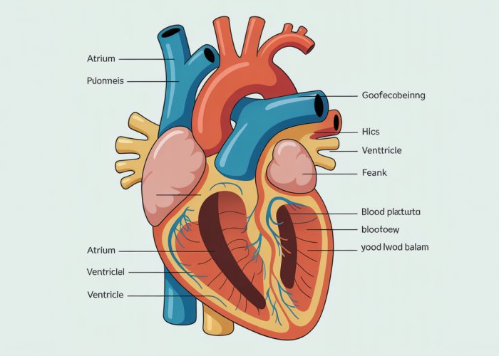
A Deep Dive into the Two-Chambered Heart Anatomy
The fish heart, while simpler than its mammalian counterpart, is a marvel of evolutionary engineering. Its two-chambered design, consisting of an atrium and a ventricle, is perfectly suited for the demands of single circulation.
But the two main chambers are not the whole story, as the sinus venosus and bulbus arteriosus play crucial roles in the circulatory cycle.
The Atrium: Gateway for Deoxygenated Blood
The atrium serves as the primary receiving chamber for deoxygenated blood returning from the body.
Its thin walls and elastic nature allow it to expand and accommodate a large volume of blood.
The atrium’s contraction initiates the cardiac cycle, gently pushing blood into the ventricle.
The Ventricle: The Pumping Powerhouse
The ventricle, with its thick muscular walls, is the heart’s main pumping chamber.
Its powerful contractions generate the pressure needed to propel blood through the gills and into the systemic circulation.
The ventricle’s robust structure ensures efficient blood delivery despite the relatively low pressure inherent in single circulation.
The Sinus Venosus: A Reservoir Role
Before blood enters the atrium, it passes through the sinus venosus.
This thin-walled sac acts as a reservoir, collecting deoxygenated blood from the body’s veins.
The sinus venosus helps to smooth blood flow into the atrium, ensuring a steady supply for the ventricle.
The Bulbus Arteriosus: Damping Pressure
Located downstream of the ventricle, the bulbus arteriosus is a large, elastic vessel.
Its primary function is to dampen the pulsatile pressure generated by the ventricle’s contractions.
By smoothing out the blood flow, the bulbus arteriosus protects the delicate gill capillaries from damage caused by high-pressure surges.
The Conus Arteriosus: Regulating Flow (In Some Fish)
In some fish species, a conus arteriosus is present between the ventricle and the bulbus arteriosus.
This structure contains valves that help to regulate blood flow to the gills.
The valves prevent backflow and ensure that blood moves in a unidirectional manner, maximizing oxygen uptake. The presence or absence of the conus arteriosus can vary depending on the species of fish.
The Gills: Where Oxygenation Happens
Having explored the architecture of the fish heart and its associated structures, the focus shifts to the crucial site where deoxygenated blood is revitalized: the gills. It is within these intricate structures that the life-sustaining process of gas exchange takes place, transforming blood depleted of oxygen into a fuel-rich elixir ready to nourish the fish’s tissues.
Understanding Gill Circulation
The gills represent an evolutionary masterpiece, perfectly adapted for extracting dissolved oxygen from the aquatic environment. Gill circulation is a highly efficient system optimized for maximizing gas exchange between the fish’s blood and the surrounding water. The entire process hinges on a delicate interplay of anatomical features and physiological mechanisms.
Deoxygenated blood, having traveled through the fish’s body, arrives at the gills via afferent branchial arteries. These arteries branch into smaller vessels, ultimately leading to the gill filaments. Gill filaments are thin, highly vascularized structures that project from the gill arches, forming the primary site of gas exchange.
The Process of Gas Exchange
The gill filaments are covered with lamellae, which are thin, plate-like structures arranged perpendicularly to the flow of water. This arrangement maximizes the surface area available for gas exchange, ensuring efficient oxygen uptake. Water flows over the lamellae in a direction opposite to the flow of blood within the capillaries. This countercurrent exchange mechanism is a critical adaptation.
Countercurrent exchange maintains a concentration gradient along the entire length of the lamellae. Blood with a lower oxygen concentration always encounters water with a higher oxygen concentration, facilitating the continuous diffusion of oxygen into the blood. Carbon dioxide, a waste product of metabolism, diffuses in the opposite direction, from the blood into the water.
The efficiency of gas exchange in the gills is also enhanced by the thinness of the lamellae. The short diffusion distance allows for rapid movement of oxygen and carbon dioxide across the respiratory membrane. Once oxygenated, blood flows from the gills via efferent branchial arteries, which converge to form the dorsal aorta.
Importance of Oxygenated Blood for Metabolism
Oxygenated blood delivered from the gills is the lifeblood of the fish, fueling all metabolic processes. The circulatory system distributes this oxygen-rich blood to every cell in the fish’s body, where it is used in cellular respiration.
Cellular respiration is the process by which cells convert glucose and oxygen into energy, producing carbon dioxide and water as byproducts. This energy is essential for all cellular functions, including muscle contraction, nerve impulse transmission, and protein synthesis.
Without a constant supply of oxygenated blood, the fish’s metabolism would grind to a halt, leading to cellular dysfunction and ultimately, death. The efficiency of gill circulation and gas exchange is, therefore, directly linked to the fish’s ability to survive and thrive in its aquatic environment. The delivery of oxygen is important for:
- Energy Production: Driving ATP synthesis, powering cellular activities.
- Waste Removal: Facilitating the removal of carbon dioxide.
- Maintaining Homeostasis: Supporting overall physiological balance.
Water flows over the lamellae in a direction opposite to the flow of blood within the capillaries. This countercurrent exchange mechanism is a critical adaptation, maximizing oxygen uptake. But even with this remarkable system in place, the efficiency of gas exchange would be severely compromised without a mechanism to ensure that blood flows in a single, forward direction. This is where the crucial role of heart valves comes into play.
Heart Valves: Ensuring Unidirectional Blood Flow
The fish heart, despite its relatively simple two-chambered structure, relies on a sophisticated system of valves to maintain the essential unidirectional flow of blood. These valves act as one-way gates, opening to allow blood to pass through and then closing to prevent backflow. This function is paramount for efficient circulation and, ultimately, for the fish’s survival.
The Importance of Unidirectional Blood Flow
Imagine a pump that allows fluid to flow both forward and backward. The efficiency of such a pump would be drastically reduced, as much of its effort would be wasted simply sloshing fluid back and forth. The same principle applies to the fish heart.
If blood were allowed to flow backward, the heart would have to work much harder to circulate the same volume of blood, placing undue stress on the organ and reducing the amount of oxygen delivered to the tissues. Unidirectional blood flow ensures that oxygenated blood is efficiently transported from the gills to the rest of the body, maximizing the benefits of gas exchange.
Mechanisms Preventing Backflow
The valves within the fish heart are strategically positioned to prevent backflow at key points in the circulatory pathway. These valves are typically flap-like structures composed of thin, flexible tissue.
When blood attempts to flow backward, the pressure forces these flaps closed, effectively sealing off the passage. The precise structure and number of valves can vary slightly between different fish species, but the fundamental principle remains the same: to ensure that blood moves only in the intended direction.
The sinus venosus, which collects deoxygenated blood, often possesses a valve that prevents backflow into the venous system. Similarly, a valve located between the atrium and the ventricle ensures that blood flows only from the atrium into the ventricle, not the other way around.
The conus arteriosus, present in some fish species, contains a series of valves that further regulate blood flow as it exits the ventricle and enters the afferent branchial arteries leading to the gills. These valves work in concert to maintain a smooth and continuous flow of blood to the gills, optimizing oxygen uptake.
The functionality of these valves is of utmost importance for the fish’s health. If valves are damaged or incompetent, blood regurgitates through the orifices. The effect of this regurgitation makes the heart work harder in order to continue to pump blood.
Frequently Asked Questions About Fish Heart Structure
This FAQ section answers some common questions about the anatomy and function of a fish heart. We hope it clarifies any points from our comprehensive guide.
How does a fish heart differ from a human heart?
Unlike the four-chambered heart of mammals, a fish heart typically has two chambers: one atrium and one ventricle. This simpler structure is sufficient for their single-loop circulatory system. Understanding this difference is key to grasping fish heart structure.
What is the conus arteriosus, and what does it do?
The conus arteriosus is a muscular outflow tract present in some fish hearts. It helps regulate blood flow and smooth out pressure pulses as blood leaves the ventricle. Not all fish species possess a conus arteriosus in their fish heart structure.
How does deoxygenated blood enter a fish heart?
Deoxygenated blood returns to the heart through the sinus venosus, a thin-walled sac that collects blood from the veins. The blood then flows into the atrium, the first chamber of the fish heart structure.
Why is the fish heart considered less efficient than a mammalian heart?
Due to its two-chambered design, the fish heart doesn’t completely separate oxygenated and deoxygenated blood. This single-loop circulation means blood pressure is lower compared to the double-loop circulation of mammals. While seemingly less efficient, the fish heart structure is perfectly adapted to the fish’s metabolic needs.
Alright, hope you’ve learned something new about fish heart structure! Now you’re one step closer to understanding these amazing creatures. Go on and explore!