The lamina dura of teeth, a dense bone layer visible on dental radiographs, plays a crucial role in tooth support. Its appearance can be indicative of periodontal health, reflecting the integrity of the alveolar bone. Changes in its density or uniformity may signal underlying conditions, prompting further investigation by dental professionals. Consequently, understanding the clinical significance of the lamina dura of teeth is essential for comprehensive oral health assessment.
Our teeth, seemingly isolated entities, are in reality integral components of a complex and interconnected system.
They don’t stand alone; rather, they are supported by a sophisticated network of structures, including the gums (gingiva), periodontal ligament, cementum, and alveolar bone.
Within this supporting cast, the alveolar bone, the bone surrounding the roots of the teeth, plays a crucial role. And within the alveolar bone resides a key player often overlooked: the lamina dura.
What is the Lamina Dura?
The lamina dura is a thin, radiopaque (meaning it appears bright on X-rays) layer of dense cortical bone that lines the tooth socket (alveolus). It essentially forms the bony wall immediately adjacent to the tooth root.
Think of it as the innermost layer of the alveolar bone, directly embracing and protecting the tooth.
It is composed of compact bone, giving it its characteristic density and providing a firm anchor for the periodontal ligament fibers that attach the tooth to the bone.
A Diagnostic Window: The Lamina Dura’s Significance
The lamina dura is far more than just a structural element. It serves as a valuable diagnostic indicator of overall dental and systemic health.
Its appearance on dental X-rays, or radiographs, can provide crucial insights into the condition of the surrounding bone and even alert dentists to potential underlying medical conditions.
Changes in its thickness, density, or continuity can signal a variety of issues, ranging from localized dental problems to systemic diseases affecting bone metabolism.
Thesis Statement: Illuminating Systemic Health Through Radiographs
The Lamina Dura’s appearance on dental X-rays (radiographs) can provide valuable insights into overall dental and systemic health.
It serves as an early warning system, alerting dentists to potential underlying issues like osteoporosis, hyperparathyroidism, or periodontal disease, long before more obvious symptoms manifest. By carefully examining the lamina dura, dentists can play a critical role in early detection and intervention, ultimately improving patient outcomes.
Our teeth, seemingly isolated entities, are in reality integral components of a complex and interconnected system.
They don’t stand alone; rather, they are supported by a sophisticated network of structures, including the gums (gingiva), periodontal ligament, cementum, and alveolar bone.
Within this supporting cast, the alveolar bone, the bone surrounding the roots of the teeth, plays a crucial role. And within the alveolar bone resides a key player often overlooked: the lamina dura.
This brings us to a deeper understanding of the lamina dura itself. Beyond its role as a diagnostic marker, the lamina dura possesses a distinct anatomy and physiology that are essential to its function and overall health.
Anatomy and Physiology: Unveiling the Lamina Dura’s Structure and Function
The lamina dura, while appearing as a simple line on a radiograph, is in fact a dynamic and specialized structure.
Its unique composition and function are intricately linked to the health and stability of the teeth and the surrounding alveolar bone.
Understanding its anatomy and physiology is crucial to appreciating its diagnostic significance.
Composition: A Fortress of Compact Bone
The lamina dura is primarily composed of dense cortical bone, also known as compact bone.
This type of bone is characterized by its tightly packed mineral matrix, making it exceptionally strong and resistant to resorption.
The high mineral content is what gives the lamina dura its radiopaque appearance on X-rays.
This compact structure provides a rigid and protective socket for the tooth root.
Function: Anchoring and Protecting the Tooth
The primary function of the lamina dura is to provide a firm anchor for the periodontal ligament (PDL).
The PDL is a network of collagen fibers that connect the tooth root to the alveolar bone.
These fibers embed into both the cementum of the tooth and the lamina dura, effectively suspending the tooth within its socket.
This arrangement allows the tooth to withstand the forces of chewing and other occlusal stresses.
The lamina dura also serves as a protective barrier for the underlying bone marrow and other sensitive tissues within the alveolar bone.
Dynamic Nature: A Reflection of Oral Health
Unlike inert bone, the lamina dura is a highly dynamic structure that constantly undergoes remodeling in response to changes in the oral environment.
This remodeling process is influenced by factors such as:
- Occlusal forces
- Inflammation
- Systemic diseases affecting bone metabolism
For example, increased occlusal forces, such as those experienced during bruxism (teeth grinding), can stimulate bone deposition, resulting in a thicker and denser lamina dura.
Conversely, inflammation associated with periodontal disease can lead to bone resorption, resulting in a thinner or discontinuous lamina dura.
Changes in bone density due to conditions like osteoporosis can also affect the lamina dura’s appearance.
In these cases, the lamina dura may appear less dense and less well-defined on radiographs.
This dynamic nature makes the lamina dura a sensitive indicator of both local and systemic factors affecting bone health.
Seeing is Believing: Dental X-Rays and Lamina Dura Visualization
The anatomical intricacies and physiological processes that govern the lamina dura’s health ultimately manifest in its radiographic appearance.
Therefore, the ability to visualize and interpret the lamina dura on dental X-rays is paramount for dentists.
By carefully examining radiographs, clinicians can glean valuable insights into the state of the alveolar bone, the health of the teeth, and even potential systemic conditions.
The Power of Radiography: Illuminating the Lamina Dura
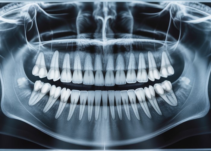
Dental X-rays, or radiographs, serve as the primary tool for visualizing the lamina dura.
These images, created by passing controlled X-ray beams through the teeth and surrounding structures, capture variations in tissue density.
Denser tissues, like bone, absorb more radiation and appear radiopaque (lighter) on the resulting image, while less dense tissues appear radiolucent (darker).
The lamina dura, due to its high mineral content and compact bone structure, exhibits a distinct radiopaque line that closely outlines the tooth root.
Different types of radiographs, such as periapical and panoramic X-rays, offer varying perspectives and levels of detail.
Periapical X-rays focus on individual teeth and their surrounding structures, providing a close-up view of the lamina dura.
Panoramic X-rays, on the other hand, capture a broader view of the entire dentition and jaw, allowing for assessment of the lamina dura’s overall continuity and integrity throughout the mouth.
A Picture of Health: The Ideal Lamina Dura
A healthy lamina dura presents as a continuous, unbroken, and uniformly radiopaque line surrounding the tooth root.
Its thickness is generally consistent, though slight variations can occur depending on the tooth’s location and function.
The presence of a well-defined lamina dura is a strong indicator of healthy bone metabolism and adequate support for the tooth.
The adjacent periodontal ligament space, appearing as a thin radiolucent line between the lamina dura and the tooth root, should also be of uniform width.
This space allows for slight tooth movement and acts as a shock absorber during chewing.
When the Image Falters: Signs of Compromise
The appearance of the lamina dura can change significantly in the presence of various pathological conditions.
These changes can manifest as alterations in its density, thickness, or continuity, offering valuable clues to the underlying problem.
Here are some examples:
-
Thinning or Disappearance: A lamina dura that appears thinner than normal or is completely absent in certain areas may indicate bone resorption.
This can be a sign of periodontal disease, osteoporosis, or other conditions that affect bone density. -
Widening: While less common, widening of the lamina dura can occur in certain situations, such as in cases of excessive occlusal forces or trauma.
-
Loss of Definition: A lamina dura that appears blurred or indistinct may suggest early bone loss or other subtle changes in bone metabolism.
-
Radiolucent Lesions: The presence of radiolucent areas adjacent to the lamina dura can indicate periapical lesions, cysts, or tumors that are eroding the surrounding bone.
It’s important to note that radiographic findings should always be interpreted in conjunction with a thorough clinical examination and patient history.
The appearance of the lamina dura is just one piece of the puzzle, and a definitive diagnosis should never be based solely on radiographic evidence.
Decoding the Lamina Dura: Health Conditions and Their Impact
While a healthy lamina dura signifies a stable and robust alveolar bone, alterations in its appearance can serve as red flags, alerting clinicians to potential underlying health conditions. These conditions range from localized oral diseases to systemic disorders affecting bone metabolism. Understanding the specific changes associated with each condition is crucial for accurate diagnosis and effective patient management.
Periodontal Disease and the Lamina Dura
Periodontal disease, an inflammatory condition affecting the tissues surrounding the teeth, can have a profound impact on the lamina dura. Chronic inflammation and infection lead to the progressive destruction of the alveolar bone, including the lamina dura.
As periodontal disease advances, the integrity of the lamina dura is compromised. It may appear blurred, discontinuous, or even absent in areas affected by bone loss. The radiographic appearance reflects the severity of the bone destruction, with more advanced cases showing significant loss of lamina dura definition.
Radiographic Appearance in Periodontitis
In cases of periodontitis, dental X-rays often reveal a loss of alveolar bone height, which directly impacts the lamina dura. The normally sharp and well-defined lamina dura may appear:
-
Fuzzy or indistinct: The clear, radiopaque line becomes less defined.
-
Discontinuous: Breaks or interruptions appear in the lamina dura.
-
Absent: In severe cases, the lamina dura may completely disappear in affected areas.
The presence of vertical bone defects, also known as infrabony pockets, alongside these changes in the lamina dura, further supports a diagnosis of periodontitis. These defects appear as angular radiolucencies adjacent to the tooth root, indicating localized bone loss.
Osteoporosis, Bone Density, and the Lamina Dura
Osteoporosis, a systemic skeletal disease characterized by low bone mass and microarchitectural deterioration of bone tissue, also affects the alveolar bone and, consequently, the lamina dura. Although dental X-rays are not designed for diagnosing osteoporosis, they can sometimes provide an early indication of reduced bone density.
The link between osteoporosis and the lamina dura lies in the overall decrease in bone mineral density. As bone becomes less dense, the lamina dura may appear thinner and less radiopaque on radiographs.
Dental X-Rays as an Early Indicator
While dual-energy X-ray absorptiometry (DEXA) scans are the gold standard for diagnosing osteoporosis, dental X-rays can sometimes offer a subtle clue. If a dentist observes a generalized thinning of the lamina dura in conjunction with other risk factors for osteoporosis (such as age, family history, or certain medications), they may recommend that the patient undergo further evaluation by a physician.
It’s important to note that dental X-rays are not a substitute for DEXA scans, and a normal-appearing lamina dura does not rule out the possibility of osteoporosis. However, suspicious radiographic findings can prompt timely intervention and potentially prevent more serious complications.
Hyperparathyroidism and its Effects
Hyperparathyroidism, a condition characterized by excessive secretion of parathyroid hormone (PTH), significantly impacts calcium metabolism and bone remodeling. PTH regulates calcium levels in the blood, and when levels are too high, it can lead to increased bone resorption. This process can affect the lamina dura and other bones throughout the body.
Radiographic Findings in Hyperparathyroidism
The increased bone resorption associated with hyperparathyroidism can manifest in several radiographic changes:
-
Loss of Lamina Dura: The lamina dura may appear thin, indistinct, or even absent. This is a common finding due to the overall bone loss.
-
Generalized Osteopenia: A decrease in bone density throughout the jaws can be observed. This makes the bones appear more radiolucent.
-
"Salt and Pepper" Appearance: This refers to a mottled appearance of the bone due to areas of increased and decreased density.
-
Brown Tumors: These are radiolucent lesions that can occur in the jaws or other bones. They are not true tumors but rather areas of bone resorption and fibrous tissue replacement.
Dentists’ Role in Diagnosis
Dentists play a crucial role in identifying potential cases of hyperparathyroidism. They are often the first healthcare professionals to notice subtle changes in the lamina dura or other radiographic signs suggestive of the condition.
By carefully evaluating dental X-rays and considering the patient’s medical history, dentists can refer patients for further evaluation by an endocrinologist. This may involve blood tests to measure PTH and calcium levels, as well as imaging studies to assess bone density. Early diagnosis and treatment of hyperparathyroidism can help prevent serious complications, such as fractures and kidney stones.
Alright, the presence of vertical bone defects, also known as infrabony pockets, alongside these changes in the lamina dura, further supports a diagnosis of periodontitis. These defects appear as…
The Dentist’s Expertise: Diagnosis, Interpretation, and Management
The ability to recognize and interpret subtle changes in the lamina dura’s appearance on dental radiographs is a cornerstone of dental practice. It’s not merely about identifying a radiopaque line; it’s about understanding its significance in the broader context of a patient’s oral and systemic health. This is where the dentist’s expertise truly shines, transforming radiographic findings into actionable clinical insights.
The Indispensable Role of Regular Dental Check-ups
Regular dental check-ups, coupled with appropriate radiographic examinations, are paramount for maintaining optimal oral health and facilitating early detection of potential issues affecting the lamina dura. These routine visits allow dentists to establish a baseline understanding of each patient’s unique anatomy and bone structure, making it easier to identify subtle deviations from the norm over time.
Dental radiographs, particularly bitewing and periapical X-rays, provide a detailed view of the teeth, surrounding bone, and the lamina dura. The frequency of these radiographs should be determined on an individual basis, considering factors such as the patient’s risk for caries, periodontal disease, and other oral health conditions.
Lamina Dura as Part of Comprehensive Dental Evaluation
The interpretation of lamina dura findings is never done in isolation. Dentists meticulously integrate these observations with other clinical findings, such as probing depths, bleeding on probing, tooth mobility, and patient history, to arrive at an accurate diagnosis. The lamina dura acts as one piece of a larger diagnostic puzzle.
For example, a blurred lamina dura, coupled with deep periodontal pockets and gingival inflammation, strongly suggests the presence of periodontitis. Similarly, a generalized loss of lamina dura definition, especially in post-menopausal women, might raise suspicion for osteoporosis and warrant further investigation.
Early Detection and Intervention: A Proactive Approach
The true value of recognizing changes in the lamina dura lies in the potential for early detection and intervention. By identifying subtle radiographic signs, dentists can initiate timely treatment and preventive measures, potentially averting more severe consequences down the line.
In the case of periodontal disease, early intervention can help control inflammation, prevent further bone loss, and preserve tooth attachment. For patients with suspected osteoporosis or hyperparathyroidism, dental findings can prompt referral to a physician for appropriate medical evaluation and management.
Managing Conditions and Patient Care
Dentists also play a crucial role in managing patient care following the initial diagnosis. This often involves educating patients about their condition, explaining the importance of adherence to treatment recommendations, and monitoring their progress over time.
For example, patients undergoing treatment for osteoporosis may experience improvements in bone density, which can be reflected in subsequent radiographic examinations of the lamina dura. Close collaboration between dentists and physicians is essential to ensure coordinated and comprehensive patient care.
Lamina Dura of Teeth: Your Health Questions Answered
Here are some common questions about the lamina dura and what it tells us about your oral and overall health.
What exactly is the lamina dura of teeth?
The lamina dura is a thin, dense layer of bone that lines the tooth socket. It appears as a radiopaque (lighter) line on dental X-rays surrounding the root of the tooth. It’s essential for anchoring the tooth to the jawbone.
What does it mean if my dentist says my lamina dura is thin or missing?
A thin or missing lamina dura on an X-ray can indicate several possible issues. These include certain metabolic diseases, osteoporosis, or recent tooth extraction. It can also sometimes suggest the presence of a tumor or lesion affecting the bone. Further investigation is needed to determine the exact cause.
Can a problem with my lamina dura directly cause tooth loss?
Not directly, but problems affecting the lamina dura of teeth can weaken the tooth’s support. This weakened support can indirectly contribute to tooth mobility and eventual tooth loss if the underlying issue isn’t addressed. The lamina dura itself is a reflection of the bone health surrounding the tooth.
How is the health of the lamina dura of teeth assessed?
The lamina dura’s health is typically assessed through dental X-rays during routine check-ups. Dentists examine the thickness, density, and overall appearance of the lamina dura on the X-ray to identify any abnormalities that may warrant further investigation or treatment.
So, next time you’re at the dentist, remember the lamina dura of teeth! It’s just one more fascinating thing they’re checking to keep your smile healthy and bright.