The remarkable diversity of Protista demands a closer examination of their cellular structures. Specifically, understanding silica deposition, a process observed in diatoms, directly informs the nature of protistan cell walls. Scientists at the Marine Biological Laboratory conduct extensive research on protistan cell structure and function. Therefore, understanding what is the cell wall of the protista is crucial for classifying these organisms. Moreover, advancements in electron microscopy allow for detailed analysis of these microscopic structures. The structural polysaccharide, chitin, forms a key component in the cell walls of certain protists.
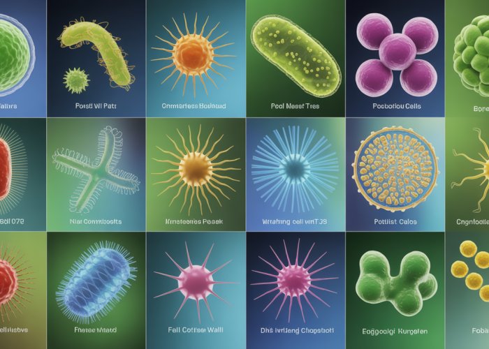
The realm of the Protista, a kingdom teeming with single-celled eukaryotic organisms, presents a captivating enigma to biologists. These microscopic entities, often overlooked, play critical roles in global ecosystems, influencing everything from nutrient cycles to the very air we breathe. A key feature defining, or rather differentiating, these diverse life forms is the presence, absence, and composition of their cell walls.
But what exactly is the cell wall of a protist? The answer, as we will explore, is far from simple.
Defining Protista: A World of Eukaryotic Diversity
Protists are fundamentally eukaryotic organisms, meaning their cells possess a nucleus and other membrane-bound organelles – a significant distinction from prokaryotic bacteria and archaea.
This eukaryotic nature places them on a more complex level of cellular organization. However, defining Protista beyond this becomes challenging due to their sheer diversity and evolutionary history. They are, in essence, a "catch-all" kingdom for eukaryotes that are not plants, animals, or fungi.
Their evolutionary significance is immense. Protists represent some of the earliest eukaryotic lineages and are considered ancestral to the other eukaryotic kingdoms. Understanding their biology provides crucial insights into the evolution of complex life on Earth.
The Cell Wall: A Universal Feature?
The cell wall, a rigid outer layer surrounding the cell membrane, is a common feature in many organisms, providing structural support, protection, and shape. However, its presence and composition vary dramatically across the biological spectrum.
In plants, the cell wall is primarily composed of cellulose. In fungi, it’s chitin. In bacteria, peptidoglycan. But what about protists?
The fascinating twist within the Protista kingdom is that the presence and composition of cell walls are far from uniform. Some protists possess rigid cell walls composed of various materials. Others have flexible outer coverings called pellicles, while still others lack any defined external structure at all.
This variability raises fundamental questions about the evolutionary pressures shaping these diverse strategies.
Exploring the Composition and Function of Protist Cell Walls
This article will delve into the composition and function of protist cell walls (or the structures that take their place), exploring the materials they are made of and the roles they play in the lives of these microorganisms.
We will examine how the presence or absence of a cell wall influences a protist’s lifestyle, ecological role, and interactions with its environment.
The central question we aim to address is: How does the structure and composition of the protist cell wall (or the alternative mechanisms of support) contribute to the incredible diversity and ecological success of this kingdom?
Acknowledging the Pioneers: Hooke and van Leeuwenhoek
Our understanding of the cellular world, including the Protista, owes a great debt to early microscopists. Robert Hooke, with his observations of cork cells in the 17th century, first coined the term "cell".
Shortly thereafter, Anton van Leeuwenhoek meticulously documented microscopic organisms, including protists, opening a window into a world previously invisible to the human eye. Their pioneering work laid the foundation for the field of microbiology and our ongoing exploration of the Protista kingdom.
What Defines a Protist? Eukaryotic Diversity and the Cell Wall Question
Having established the foundational role of the cell wall in the broader biological landscape, it’s time to turn our attention to how it manifests—or doesn’t—within the Protista. The cell wall is far from a universal characteristic of these organisms.
Eukaryotes vs. Prokaryotes: A Fundamental Divide
Protists are unequivocally eukaryotic.
This means their cells possess a defined nucleus and other membrane-bound organelles, a feature utterly absent in prokaryotic cells like bacteria and archaea.
This structural complexity allows for greater compartmentalization and efficiency in cellular processes, a hallmark of eukaryotic life.
The distinction between eukaryotes and prokaryotes is one of the most fundamental divisions in biology, and it immediately situates Protista within a specific domain of life.
The Protist Puzzle: A Classification Conundrum
Defining and classifying protists has been, and continues to be, a significant challenge in biology.
Historically, they were often described as a "catch-all" group for eukaryotes that didn’t neatly fit into the plant, animal, or fungi kingdoms.
Modern molecular phylogenetics has revealed that Protista is not a monophyletic group, meaning they don’t all share a single common ancestor.
Instead, they represent a diverse collection of evolutionary lineages, some more closely related to plants, animals, or fungi than to other protists.
This complex evolutionary history makes it difficult to establish a clear and universally accepted classification system.
The ongoing refinement of protist taxonomy highlights the dynamic nature of scientific understanding and the power of new technologies in uncovering evolutionary relationships.
Nutritional Diversity: Autotrophs and Heterotrophs
Protists exhibit a remarkable range of nutritional strategies.
Some are photosynthetic autotrophs, capable of producing their own food through photosynthesis, much like plants. Algae are prime examples.
Others are heterotrophs, obtaining nutrients by consuming other organisms or organic matter. This includes predators, parasites, and decomposers.
Still others are mixotrophs, capable of both photosynthesis and heterotrophic feeding, depending on environmental conditions.
This nutritional diversity reflects the varied ecological roles that protists play in ecosystems, from primary producers to consumers and decomposers.
Cell Wall as a Taxonomic Tool
The presence or absence of a cell wall, along with its composition, can be a valuable tool for classifying and differentiating protists.
Some protists, like certain algae, possess cell walls composed of cellulose, similar to plants.
Others, such as diatoms, have intricate cell walls made of silica, creating beautiful and durable structures.
Still others, like amoebas, lack a rigid cell wall altogether, relying on their cell membrane and cytoskeleton for support and movement.
These differences in cell wall characteristics provide clues about the evolutionary relationships between protists and their adaptations to specific environments.
Osmoregulation and Contractile Vacuoles
Protists, particularly those living in freshwater environments, face the challenge of osmoregulation: maintaining a stable internal water balance.
Because the concentration of solutes is usually higher inside the cell than in the surrounding water, water tends to enter the cell by osmosis.
To counteract this, many protists possess contractile vacuoles, organelles that actively pump excess water out of the cell.
This prevents the cell from swelling and bursting, ensuring its survival in a hypotonic environment.
The presence and function of contractile vacuoles are another important characteristic that distinguishes different groups of protists.
The Cell Wall: Structure and Function in the Microscopic Realm
As we delve deeper into the world of protists, the presence or absence of a cell wall becomes a crucial differentiating factor. However, before exploring the specifics of protist cell walls, it’s important to establish a firm understanding of what a cell wall is and what it does in biological systems.
A cell wall, in its most basic definition, is a rigid, yet often flexible, layer located outside the cell membrane. This outer layer is a fundamental component for various organisms, from plants and bacteria to fungi and, as we’ll see, many protists.
Defining the Cell Wall
The cell wall is defined as the non-living, rigid layer that surrounds the plasma membrane of cells in plants, algae, fungi, and many prokaryotes. It’s a complex structure with a remarkable ability to provide support and protection to the cell.
Unlike the cell membrane, which is flexible and dynamic, the cell wall is relatively static.
It provides a defined shape and contributes to the overall mechanical integrity of the cell.
The Multifaceted Functions of a Cell Wall
The cell wall isn’t just a passive barrier; it performs several crucial functions that are vital to a cell’s survival. These functions can be broadly categorized as:
-
Structural Support: Perhaps the most obvious function, the cell wall provides the cell with physical support, allowing it to maintain its shape and resist external forces. Without a cell wall, many cells would simply collapse.
-
Protection from Osmotic Pressure: Cells often exist in environments where the concentration of solutes differs from their internal environment. This difference in concentration creates osmotic pressure, which can cause cells to either swell and burst (in hypotonic solutions) or shrivel up (in hypertonic solutions). The cell wall acts as a counter-pressure, preventing the cell from either bursting or collapsing due to osmotic imbalances.
-
Defining Cell Shape: By providing a rigid outer boundary, the cell wall dictates the shape of the cell. This is particularly important for organisms like diatoms, where the intricate cell wall structure contributes to their characteristic and beautiful forms.
Cell Wall vs. Cell Membrane vs. Pellicle: A Comparative Overview
While the cell wall, cell membrane, and pellicle all contribute to a cell’s structure and protection, they are distinct entities with different properties.
The cell membrane is a flexible, selectively permeable barrier that regulates the passage of substances in and out of the cell. It is composed of a phospholipid bilayer with embedded proteins.
The cell wall is a rigid, non-living layer that provides structural support and protection. It is located outside the cell membrane.
The pellicle, found in some protists like Euglena, is a flexible, proteinaceous layer beneath the cell membrane that provides shape and support without the rigidity of a true cell wall.
In essence:
- The cell membrane controls what enters and exits the cell.
- The cell wall provides shape and support.
- The pellicle offers flexibility and support in the absence of a cell wall.
Understanding these distinctions is crucial for appreciating the diversity of strategies that cells employ to maintain their integrity and thrive in various environments.
Cell Wall Composition: A Tour of Protist Diversity
Having explored the multifaceted roles that cell walls play in the lives of various organisms, it’s time to consider the remarkable diversity in the chemical makeup of these structures, particularly within the protist kingdom. From sturdy polysaccharides to intricate mineral matrices, protist cell walls exhibit a fascinating array of compositions, each reflecting unique adaptations to their respective environments.
The Building Blocks of Protist Cell Walls
Unlike the relatively uniform composition seen in plant cell walls (primarily cellulose), protist cell walls showcase a far greater variety of materials. This heterogeneity reflects the evolutionary distance between different protist lineages and the diverse ecological niches they occupy.
Cellulose: A Familiar Framework
Cellulose, a complex polysaccharide composed of glucose monomers, is perhaps the most recognizable component of cell walls. While famously abundant in plants, cellulose also forms the structural basis for the cell walls of certain algal protists. These algae, often found in freshwater environments, rely on the strength and rigidity of cellulose to maintain their cell shape and withstand osmotic pressures.
Silica: Glass Houses of the Microscopic World
In stark contrast to the organic nature of cellulose, some protists construct their cell walls from inorganic materials. Diatoms, a group of single-celled algae, are renowned for their intricate cell walls, known as frustules, made of silica (silicon dioxide). These glassy structures exhibit a remarkable diversity of shapes and patterns, often described as "jewels of the sea."
The porous nature of silica frustules allows for efficient nutrient uptake and waste removal.
Diatoms exploit light more efficiently because of their silica-based cell walls.
Calcium Carbonate: A Calcareous Armor
Another mineral found in protist cell walls is calcium carbonate (CaCO3). This compound, commonly associated with the shells of marine organisms, also serves as a protective barrier for certain protists, particularly those inhabiting calcium-rich marine environments. The hard, chalky nature of calcium carbonate provides a robust defense against predators and physical stressors.
Composition, Environment, and Lifestyle
The specific composition of a protist’s cell wall is intricately linked to its environment and lifestyle.
For instance, diatoms with their silica frustules thrive in aquatic environments.
These environments have readily available dissolved silica.
Cellulose-based cell walls in freshwater algae provide structural support in hypotonic environments, where water tends to flow into the cell.
Calcium carbonate cell walls are advantageous in marine environments.
Here, they offer protection from predation and contribute to the formation of marine sediments.
Ecological Significance
Protist cell wall composition isn’t merely a matter of structural integrity; it also plays a crucial role in the ecological functions of these organisms.
Diatoms, with their silica frustules, are responsible for a significant portion of global carbon fixation.
When they die, their silica shells sink to the ocean floor, effectively sequestering carbon.
Calcium carbonate-shelled protists also contribute to carbon cycling and the formation of limestone deposits.
Cellulose-walled algae form the base of many aquatic food webs.
Their photosynthetic activity supports a diverse range of organisms.
The diverse array of materials found in protist cell walls underscores the remarkable adaptability of these microorganisms. Each composition reflects a unique set of selective pressures and contributes to the ecological roles these organisms play in their respective environments.
Protists Without Walls: The Pellicle and Other Adaptations
We’ve seen how diverse the composition of protist cell walls can be, from the cellulose of some algae to the silica frustules of diatoms. But what about protists that forgo the traditional cell wall altogether? These organisms have evolved alternative strategies for maintaining their shape, protecting themselves, and navigating their environments. One notable example is the pellicle, a structure that offers a unique blend of support and flexibility.
The Case of the Missing Cell Wall
The absence of a rigid cell wall might seem like a disadvantage. However, for certain protists, it provides significant adaptive benefits. These organisms, often found in dynamic or nutrient-rich environments, rely on alternative support systems. This allows for greater motility and adaptability.
The Pellicle: A Flexible Fortress
The pellicle is a specialized structure found in some protists, most notably Euglena and trypanosomes. It lies directly beneath the cell membrane and consists of a series of interlocking protein strips.
These strips are often supported by microtubules, providing structural integrity while still allowing for cellular flexibility and movement. The pellicle enables these protists to change shape, squeeze through tight spaces, and exhibit a characteristic wriggling motion known as metaboly.
Structure of the Pellicle
The pellicle isn’t a single, monolithic structure, but rather a complex assembly of proteins.
- Protein Strips: These form the main supporting framework, arranged in a spiral pattern around the cell.
- Microtubules: These run parallel to the protein strips, providing additional support and helping to maintain cell shape.
- Cell Membrane: This encloses the pellicle, acting as the outermost boundary of the cell.
Functionality: More Than Just Support
Beyond structural support, the pellicle plays several other important roles:
- Shape Maintenance: It helps the protist maintain a recognizable, albeit flexible, shape.
- Motility: It facilitates movement through metaboly, allowing the protist to squeeze and contort its body.
- Protection: While not as robust as a cell wall, the pellicle offers a degree of protection against physical stress and osmotic changes.
Pellicle vs. Cell Wall: A Comparative Glance
| Feature | Pellicle | Cell Wall |
|---|---|---|
| Rigidity | Flexible | Rigid |
| Composition | Protein strips, microtubules | Varies (cellulose, silica, calcium carbonate, etc.) |
| Function | Support, motility, limited protection | Support, protection, shape |
| Organisms | Euglena, trypanosomes | Algae, diatoms, some bacteria |
Advantages and Disadvantages
Advantages of the Pellicle:
- Flexibility: Allows for movement through tight spaces and shape changes.
- Metaboly: Enables a unique form of locomotion.
- Adaptability: Suits organisms in dynamic environments.
Disadvantages of the Pellicle:
- Limited Protection: Offers less protection than a rigid cell wall.
- Osmotic Sensitivity: Pellicle-bearing protists are often more sensitive to osmotic changes.
- Structural Limitations: May not be suitable for large cells or those requiring strong structural support.
In conclusion, the pellicle represents a fascinating adaptation in protists that lack a traditional cell wall. Its unique structure and function allow these organisms to thrive in diverse environments, showcasing the remarkable evolutionary solutions found within the microscopic world. While it may lack the robust protection of a cell wall, the pellicle provides a compelling alternative, balancing support with the flexibility needed for survival.
Diatoms: Jewels of the Microscopic World and Their Silica Cell Walls
Having considered protists that cleverly circumvent the need for a rigid wall, it’s time to turn our attention to organisms that embrace cell wall architecture with extraordinary artistry. Among these, diatoms stand out, not just for possessing cell walls, but for the sheer beauty and functional elegance of their glassy fortresses.
Introducing Diatoms and Their Frustules
Diatoms are a group of single-celled algae renowned for their unique silica cell walls, known as frustules. These microscopic marvels are essentially tiny, ornate glass houses that encase the diatom cell. They are found in virtually all aquatic environments, from oceans and lakes to rivers and even moist soil.
Unlike the cellulose walls of many other algae, diatom frustules are made of biogenic silica, a hydrated form of silicon dioxide (SiO₂·nH₂O). This material gives the frustules their characteristic transparency and glass-like appearance.
Structure and Function of Diatom Frustules
The frustule is composed of two overlapping halves, or valves: the epitheca (the larger, upper valve) and the hypotheca (the smaller, lower valve). These valves fit together much like a petri dish, creating a protective and intricately patterned shell.
The surface of the frustule is adorned with a variety of pores, grooves, and ridges, which are not merely decorative but serve important functions.
These features enhance surface area for nutrient uptake and gas exchange. They also provide structural support and may play a role in buoyancy and light diffusion.
The intricate patterns on diatom frustules are species-specific, making them invaluable tools for taxonomic identification.
The silica composition provides considerable rigidity and protection from predation, while the intricate patterns likely contribute to optimized light capture for photosynthesis.
Ecological Significance of Diatoms
Diatoms are primary producers, meaning they convert sunlight into energy through photosynthesis. They are responsible for an estimated 20% of global oxygen production, rivaling rainforests in their contribution to the Earth’s atmosphere.
Diatoms form the base of many aquatic food webs, serving as a crucial food source for zooplankton and other organisms.
Furthermore, diatoms play a significant role in the global carbon cycle. As they photosynthesize, they absorb carbon dioxide from the atmosphere and incorporate it into their biomass.
When diatoms die, their silica frustules sink to the ocean floor, effectively sequestering carbon for long periods. This process, known as the biological carbon pump, helps regulate the Earth’s climate.
Diatomaceous earth, a sedimentary deposit composed of fossilized diatom frustules, has numerous industrial applications, including filtration, insulation, and abrasives.
Microscopy and the Study of Diatom Cell Walls
The intricate details of diatom frustules can only be fully appreciated through microscopy. Light microscopy, particularly phase contrast microscopy, is commonly used to observe the overall structure and patterns of frustules.
However, electron microscopy (both scanning electron microscopy or SEM, and transmission electron microscopy or TEM) provides much higher resolution. This allows for the examination of the fine details of the silica structure, including the size and arrangement of pores and other surface features.
Microscopy not only allows scientists to study the morphology of diatoms. It also helps them understand the biomineralization processes involved in frustule formation, and the evolutionary relationships between different diatom species. Advanced techniques, such as atomic force microscopy (AFM), can even be used to probe the mechanical properties of the frustules.
The Ecological Significance of Protist Cell Walls
From the vast expanse of the oceans to the hidden depths of soil, protists are ubiquitous. Their cell walls, diverse in composition and structure, play a critical role in shaping the ecology of these diverse ecosystems.
They actively engage in nutrient cycles and influence global biogeochemical processes. The cell walls of these microscopic organisms are not merely protective barriers; they are integral components of the planet’s life support systems.
Nutrient Cycling: A Foundation of Ecosystem Health
Protist cell walls are crucial to the cycling of essential nutrients. After a protist dies, the cell wall is broken down by bacteria and fungi, returning its constituent elements to the environment.
Consider the silica frustules of diatoms. As these organisms die, their silica shells sink to the ocean floor, forming diatomaceous earth deposits.
These deposits act as a long-term sink for silicon, an element vital for the growth of other organisms. This process ensures the continued availability of this nutrient within the marine ecosystem.
In freshwater environments, the cell walls of algae like desmids, composed primarily of cellulose, contribute to the carbon cycle. Their decomposition releases carbon back into the water, fueling the growth of other microorganisms and aquatic plants.
Diatoms and Carbon Sequestration: A Climate Change Ally
Diatoms are responsible for an estimated 20% of global photosynthesis. Their silica cell walls are central to their role in carbon sequestration.
During photosynthesis, diatoms absorb carbon dioxide from the atmosphere and convert it into organic matter. When these diatoms die, their silica frustules, encasing the carbon-rich organic material, sink to the ocean floor.
This process effectively removes carbon from the atmosphere and stores it in the deep ocean for extended periods. This natural carbon sequestration mechanism is a crucial component of the global carbon cycle and helps regulate Earth’s climate.
The efficiency of this process is directly linked to the structural integrity and sinking rate of diatom frustules. Any disruption to diatom populations or the formation of their silica cell walls could have significant implications for climate change.
Bioremediation Potential: Harnessing Protist Power
The unique properties of protist cell walls hold promise for bioremediation, the use of biological organisms to remove pollutants from the environment. Certain protists can accumulate heavy metals or other contaminants within their cell walls.
For example, some algae species can absorb heavy metals like lead and cadmium, effectively removing them from contaminated water. The cell walls act as a biosorbent, binding to the pollutants and preventing them from spreading further.
After the protists have accumulated the pollutants, they can be harvested, and the contaminants safely disposed of or even recycled.
This approach offers a sustainable and cost-effective alternative to traditional chemical methods of remediation. However, further research is needed to fully explore and optimize the bioremediation potential of protist cell walls.
Climate Change Impacts on Protist Cell Walls
Climate change poses a significant threat to protist populations and the formation of their cell walls. Ocean acidification, caused by increased atmospheric carbon dioxide, can affect the ability of diatoms and other protists to produce their silica or calcium carbonate shells.
Warmer water temperatures can also alter the distribution and abundance of protist species. This change can disrupt marine food webs and impact carbon cycling.
Changes in salinity and nutrient availability can also affect the structure and composition of protist cell walls, potentially altering their ecological functions. Understanding these impacts is crucial for predicting how climate change will affect marine ecosystems and the global carbon cycle.
Protist Cell Walls and Disease: When Microbes Turn Menacing
As we’ve seen, protist cell walls play vital roles in ecology, but their influence extends beyond these beneficial functions. In some cases, the very features that allow protists to thrive can also make them agents of disease. Understanding how cell walls contribute to protist pathogenicity is crucial for developing effective treatments against these microscopic invaders.
Pathogenic Protists: A Rogues’ Gallery
Several protists are notorious for causing diseases in humans and other animals. Plasmodium, the causative agent of malaria, is perhaps the most well-known.
Others include Trypanosoma, responsible for diseases like sleeping sickness and Chagas disease, and Giardia, a common cause of intestinal infections. Entamoeba histolytica is another significant pathogen, causing amoebic dysentery and liver abscesses.
The Cell Wall’s Role in Pathogenicity
The cell wall, or lack thereof, significantly impacts a protist’s ability to cause disease. Protists like Plasmodium and Trypanosoma lack a conventional cell wall, instead possessing a flexible pellicle or a surface coat of glycoproteins.
This structural characteristic aids in immune evasion and facilitates their complex life cycles within different hosts and vectors. The absence of a rigid cell wall allows for rapid shape changes, which can be beneficial for penetrating host cells or evading immune responses.
Giardia’s Tough Cyst Wall: A Barrier to Treatment
In contrast, protists like Giardia possess a tough, resistant cyst wall. This wall protects the parasite during its transmission stage, allowing it to survive harsh environmental conditions outside the host.
The cyst wall is composed of chitin and other polysaccharides, making it highly resistant to degradation and disinfection. This resilience is a major factor in the persistence and widespread transmission of giardiasis, as the cysts can survive for extended periods in water sources.
Entamoeba histolytica and Cell Wall Lysis
Entamoeba histolytica, while lacking a defined cell wall in its trophozoite form, relies on its ability to secrete enzymes that degrade the extracellular matrix and cell walls of host tissues. This allows the amoeba to invade the intestinal lining, causing ulcers and dysentery.
The absence of a rigid cell wall in the trophozoite stage facilitates this invasive process, enabling the parasite to move through tissues and access nutrients.
Targeting Cell Wall Structures for Treatment
Understanding the structure and composition of protist cell walls, or the surface structures that take their place, opens avenues for developing targeted treatments.
For instance, drugs that disrupt the synthesis or assembly of the cyst wall in Giardia could prevent the formation of infective cysts, reducing transmission. Similarly, research into the surface glycoproteins of Trypanosoma could lead to the development of vaccines or therapies that block the parasite’s ability to evade the immune system.
In the case of Entamoeba histolytica, understanding the mechanisms by which the amoeba degrades host tissues could lead to the development of inhibitors that prevent tissue invasion.
By focusing on the unique structural features of pathogenic protists, researchers can design more effective and specific treatments, ultimately reducing the burden of these debilitating diseases.
The cyst walls of pathogens such as Giardia are notoriously difficult to penetrate, hindering the effectiveness of many treatments. But how do scientists unravel the intricacies of these microscopic defenses? The answer lies in the powerful realm of microscopy, a field that has revolutionized our understanding of protist cell walls and their multifaceted roles.
Microscopy and the Exploration of Protist Cell Walls
Microscopy serves as an indispensable tool for unveiling the hidden world of protist cell walls. These structures, often too small to be seen with the naked eye, become accessible through the magnifying power of various microscopic techniques.
Microscopy enables researchers to examine the structural details, compositional elements, and functional properties of these walls. This allows them to understand the diverse roles protist cell walls play in ecology, disease, and other biological processes.
The Power of Visualization: Microscopy Unveiled
Microscopy is fundamental to the study of protist cell walls because it allows us to visualize structures that would otherwise remain invisible. The resolution and magnification offered by different microscopy techniques allow scientists to discern the intricate details of cell wall architecture.
This visualization is essential for understanding how these structures contribute to a protist’s survival, its interactions with the environment, and its potential to cause disease.
A Toolkit of Techniques: Exploring Different Microscopy Methods
Numerous microscopy techniques are employed to study protist cell walls, each offering unique advantages and insights.
The most common methods include light microscopy and electron microscopy, but other specialized techniques are also used. Each method brings a unique perspective to studying these microscopic structures.
Light Microscopy: A Foundation for Discovery
Light microscopy, including brightfield, phase contrast, and fluorescence microscopy, has been instrumental in the initial characterization of protist cell walls.
These techniques are relatively simple and inexpensive, making them widely accessible to researchers. Light microscopy allows for the observation of live cells and the visualization of cell wall structures under various conditions. Staining techniques can further enhance the contrast and reveal specific components within the cell wall.
Electron Microscopy: Unveiling Ultrastructural Details
Electron microscopy provides a higher resolution than light microscopy, enabling the visualization of the ultrastructure of protist cell walls.
There are two main types of electron microscopy: transmission electron microscopy (TEM) and scanning electron microscopy (SEM).
-
Transmission Electron Microscopy (TEM): TEM involves passing a beam of electrons through a thin section of the sample. This technique reveals the internal structure and organization of the cell wall at the nanometer scale. TEM is invaluable for studying the layers, pores, and other fine details of the cell wall.
-
Scanning Electron Microscopy (SEM): SEM involves scanning the surface of the sample with a focused beam of electrons. This technique provides a three-dimensional view of the cell wall’s surface topography. SEM is particularly useful for examining the external features, such as the intricate patterns on diatom frustules.
What Microscopy Reveals: Structure and Composition in Detail
Microscopy provides critical information about both the structure and composition of protist cell walls. The specific details revealed depend on the microscopy technique used and the sample preparation methods.
Structural Insights: From Layers to Pores
Microscopy allows researchers to examine the layering, thickness, and organization of cell walls.
For example, electron microscopy can reveal the distinct layers in the cell walls of certain algae or the complex pore structures in diatom frustules. Understanding these structural features is essential for determining how the cell wall functions in protection, support, and interaction with the environment.
Compositional Analysis: Identifying Key Components
While microscopy primarily provides structural information, it can also be combined with staining or labeling techniques to identify specific components within the cell wall.
For instance, fluorescently labeled antibodies can be used to detect the presence of specific proteins or polysaccharides in the cell wall. Spectroscopic techniques, such as energy-dispersive X-ray spectroscopy (EDS) coupled with electron microscopy, can also be used to determine the elemental composition of the cell wall. This allows scientists to identify the presence of silica, calcium carbonate, or other materials.
Frequently Asked Questions: Protista Cell Walls
Here are some common questions about protista cell walls to help you better understand these microscopic structures.
What exactly is a cell wall, and do all protists have one?
A cell wall is a structural layer external to the cell membrane, providing support and protection. Not all protists possess a cell wall. Some, like amoebas, are flexible and lack a rigid wall.
What is the cell wall of the protista composed of if not cellulose?
While plant cell walls are primarily made of cellulose, protista cell walls have diverse compositions. Some contain silica (like diatoms), others calcium carbonate, and some are made of protein layers or other polysaccharides. The cell wall of the protista is highly variable depending on the species.
Why is the composition of protist cell walls so diverse?
The diversity in composition reflects the wide range of protist lifestyles and evolutionary histories. Different materials provide different types of protection or structural support suited to their specific environments.
How does the presence or absence of a cell wall affect a protist’s movement?
Protists with cell walls often move using flagella or cilia, which protrude through the wall. Protists without cell walls, like amoebas, can move by extending pseudopods, a form of cytoplasmic streaming not possible with a rigid wall.
So, that’s a peek into the fascinating world of protistan cell walls! Hopefully, you now have a better grasp of what is the cell wall of the protista and why it’s so important to these tiny organisms. Until next time, keep exploring the amazing world of microbiology!