The periosteum, the outer fibrous layer of bone, plays a critical role in bone growth and repair. Haversian canals, conversely, are intricate networks of vessels within the bone itself, facilitating nutrient delivery. Understanding how these two distinct systems communicate is central to comprehending bone physiology, where the key lies in Volkmann’s canals. Therefore, what canal connects the periosteum to the haversian canal? The answer, as understood within the field of histology, is Volkmann’s canals, the channels allowing blood vessels and nerves to transverse from the periosteum to the Haversian canals, and vice versa.
Bones are far more than just a rigid framework. They are dynamic, living tissues that provide structure, protect vital organs, and facilitate movement.
The skeletal system supports the entire body. It also serves as a crucial reservoir for essential minerals.
The Interconnected World Within Bone
Bone tissue is a complex composite of cells, a protein matrix (primarily collagen), and mineral deposits.
This intricate structure necessitates a robust system for nutrient delivery and waste removal. Without it, bone cells, like all living cells, cannot survive.
The interconnectedness of bone components is critical for its health and function.
Nutrient delivery ensures that osteocytes, the mature bone cells, receive the necessary oxygen, glucose, and other vital substances.
Concurrently, waste removal clears away metabolic byproducts, preventing their accumulation and potential toxicity.
Volkmann’s Canals: The Unsung Heroes
Deep within the bone matrix lies a network of tiny channels known as Volkmann’s canals. They are also referred to as perforating canals.
These canals serve as critical conduits. They connect the outer surface of the bone (the periosteum) to the Haversian canals, which run longitudinally within the bone.
This connection is the key to bone’s vascular and nerve supply.
Volkmann’s canals are the often-overlooked link. They allow blood vessels and nerves from the periosteum to penetrate the dense bone tissue and reach the Haversian systems.
This intricate network ensures that every bone cell receives the nourishment it needs and can effectively eliminate waste.
Article Objective
This article aims to illuminate the significance of Volkmann’s canals. We will explore their pivotal role in maintaining bone health.
By understanding the function of these canals, we can gain a deeper appreciation for the complexity and resilience of the skeletal system.
Bones are dynamic living tissues that provide support, protection, and mineral storage, but to fully appreciate the role of Volkmann’s canals, it’s essential to first delve into the fundamental architecture of bone itself.
Bone Architecture 101: Understanding the Foundation
Bone isn’t simply a solid, uniform structure. It’s a meticulously organized composite material designed for strength, flexibility, and continuous remodeling.
Understanding its components and organization is key to appreciating how Volkmann’s canals fit into the bigger picture.
The Building Blocks of Bone
At its core, bone consists of three primary components: specialized cells, an extracellular matrix, and the periosteum.
Bone cells are responsible for bone formation, maintenance, and resorption.
Bone Cells: The Workforce
- Osteoblasts are the bone-building cells. They synthesize and secrete the organic components of the bone matrix (osteoid).
- Osteocytes are mature bone cells embedded within the bone matrix (lacunae). They maintain bone tissue and act as mechanosensors.
- Osteoclasts are large, multinucleated cells responsible for bone resorption. They break down bone tissue, releasing minerals into the bloodstream.
Bone Matrix: The Structural Framework
The bone matrix is composed of both organic and inorganic materials.
- The organic component is primarily collagen. It provides flexibility and tensile strength.
- The inorganic component consists of mineral salts, primarily calcium phosphate in the form of hydroxyapatite. It provides hardness and rigidity.
This combination allows bone to withstand significant stress and pressure.
Periosteum: The Outer Layer
The periosteum is a tough, fibrous membrane covering the outer surface of bones (except at joints). It contains:
- Blood vessels
- Nerves
- Bone-forming cells
It plays a crucial role in bone growth, repair, and sensory function.
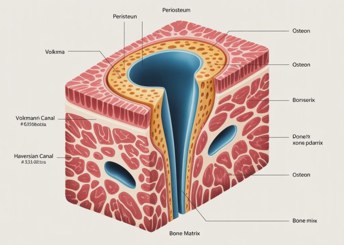
Osteons (Haversian Systems): The Functional Units
Compact bone, which forms the dense outer layer of most bones, is organized into osteons, also known as Haversian systems.
These are cylindrical structures that run parallel to the long axis of the bone.
Each osteon consists of concentric layers, or lamellae, of bone matrix surrounding a central Haversian canal.
Haversian Canals: Central Pathways
Haversian canals contain blood vessels and nerves. They supply nutrients to and remove waste from the osteocytes within the osteon.
These canals run longitudinally through the bone. They providing vital pathways for communication and sustenance.
Lacunae and Canaliculi: Microscopic Networks
Within the lamellae are small spaces called lacunae. Each lacuna houses an osteocyte.
Tiny channels called canaliculi radiate outward from the lacunae. They connect to adjacent lacunae and, ultimately, to the Haversian canal.
This intricate network allows osteocytes to exchange nutrients and waste products with the blood vessels in the Haversian canal. This is crucial for their survival and function.
The symphony of bone architecture, with its intricate cellular workforce and meticulously organized matrix, orchestrates the vital functions of support and protection. However, these internal structures rely on a crucial interface with the body’s broader systems to thrive.
The Periosteum: Bone’s Outer Protector and Nourisher
The periosteum is far more than just a superficial membrane; it’s the bone’s dynamic interface with the body, serving as its outer protector and nourisher. This resilient, fibrous sheath envelops nearly all external bone surfaces, acting as a critical mediator for growth, repair, and sensory input.
Structure and Composition
The periosteum is a two-layered structure. The outer fibrous layer provides mechanical protection, while the inner cambium layer is highly cellular and vascular. This inner layer is rich in osteoblasts, particularly during growth and fracture repair.
Collagen fibers, predominantly Type I, give the periosteum its tensile strength, anchoring it to the bone via Sharpey’s fibers that penetrate the underlying bone matrix.
This firm attachment is crucial for distributing forces and maintaining the periosteum’s integrity.
Appositional Growth: Building Bone Outward
The periosteum plays a key role in appositional bone growth, the process by which bones increase in diameter. Osteoblasts within the cambium layer deposit new bone matrix on the external surface. This process allows bones to thicken and strengthen throughout life, especially in response to increased mechanical demands.
Unlike endochondral ossification, which is responsible for longitudinal bone growth at the epiphyseal plates, appositional growth occurs directly on the existing bone surface.
Fracture Repair: The Periosteum’s Regenerative Power
When a bone fractures, the periosteum leaps into action. The blood vessels within the periosteum rupture, forming a hematoma. This initiates an inflammatory response, attracting mesenchymal stem cells to the site.
These cells differentiate into osteoblasts and chondroblasts, which begin to form a callus, a temporary bridge of cartilage and bone that stabilizes the fracture. The periosteum then contributes significantly to the remodeling of the callus into mature bone, restoring the bone’s structural integrity.
The extent of periosteal involvement in fracture healing depends on factors such as the severity of the fracture, the age of the individual, and the adequacy of blood supply.
Sensory Innervation: Feeling Bone Pain
Bones, often perceived as inert structures, are actually highly innervated. The periosteum is richly supplied with sensory nerve endings, making it highly sensitive to pain. This explains why bone fractures and injuries are so painful. The periosteal nerves transmit pain signals to the central nervous system, alerting the body to tissue damage and promoting protective behaviors.
Vascular Connection: The Lifeline to Bone
The periosteum is a highly vascularized tissue, containing a network of blood vessels that supply nutrients and oxygen to the underlying bone. These vessels penetrate the bone through Volkmann’s canals, establishing a vital connection with the Haversian canals within the bone matrix. This vascular network is essential for maintaining the viability of bone cells and supporting bone remodeling. The periosteal blood supply is also crucial for delivering immune cells and growth factors to sites of injury or infection.
Bridging the Gap: Connecting Inner and Outer Bone
The periosteum serves as a crucial bridge, not just structurally, but also functionally. The nerves and blood vessels coursing within its layers must connect with the inner bone to ensure the survival of the osteocytes and enable bone repair and growth. This connection is achieved through Volkmann’s canals, which act as perforating channels that allow these essential structures to penetrate the hard, dense bone tissue.
The periosteum, with its capacity for growth and repair, depends on a continuous exchange with the bone’s internal environment. This crucial interaction is orchestrated by a network of specialized channels. These channels act as the essential link that connects the outer surface of the bone to its innermost structures.
Volkmann’s Canals: The Vital Connection – Bridging Periosteum and Haversian Canals
At the heart of bone’s intricate vascular system lies a network of channels known as Volkmann’s Canals, also referred to as Perforating Canals.
These canals are not merely passive conduits; they are the vital connectors that bridge the gap between the periosteum and the Haversian canals. These Haversian canals reside deep within the bone’s osteons.
Defining Volkmann’s Canals
Volkmann’s canals can be defined as the microscopic channels present in bone tissue that facilitate communication between the bone’s surface and its deeper structures.
They serve as conduits for blood vessels and nerves, enabling nutrient supply and waste removal throughout the bone.
Connecting Periosteum and Haversian Canals
The primary function of Volkmann’s canals is to act as a direct link between the blood vessels and nerves present in the periosteum and those within the Haversian canals.
The Haversian canals run longitudinally through the bone, forming the central channels of the osteons. Volkmann’s canals then act as transverse channels to connect these Haversian systems to each other and to the periosteum.
This connection is critical for maintaining the health and viability of the bone tissue.
Orientation and Network Formation
Unlike Haversian canals, which run parallel to the long axis of the bone, Volkmann’s canals run perpendicular or at oblique angles. This arrangement creates an interconnected, three-dimensional network throughout the bone matrix.
This network ensures that all areas of the bone tissue are adequately supplied with nutrients and can efficiently eliminate waste products.
The branching and interconnected nature of Volkmann’s canals guarantees resilience and redundancy in the vascular supply.
Nutrient Delivery and Waste Removal
The interconnected network formed by Volkmann’s and Haversian canals is vital for the survival of osteocytes, the mature bone cells that reside within lacunae.
These lacunae are small spaces within the bone matrix, and the osteocytes within them are connected to each other and to the canals via tiny channels called canaliculi.
Through this network, oxygen, glucose, and other essential nutrients are delivered to the osteocytes. Waste products such as carbon dioxide are removed.
This constant exchange is essential for maintaining bone cell viability. It supports the overall health and structural integrity of the bone.
The intricate network of Volkmann’s canals plays a pivotal role, far beyond simply connecting different bone structures. They are, in essence, the lifelines of the bone, responsible for its continuous nourishment and the removal of metabolic byproducts. Understanding their significance is crucial to appreciating the overall health and vitality of the skeletal system.
Sustaining Bone Health: The Critical Role of Volkmann’s Canals
Volkmann’s canals are not merely structural conduits; they are fundamental to the metabolic activity that sustains bone tissue. Without their efficient function, bone would be unable to maintain its density, repair itself, or respond to the dynamic demands placed upon it.
Nutrient Delivery and Waste Removal: The Core Function
The primary responsibility of Volkmann’s canals lies in ensuring that every osteocyte, the mature bone cell, receives the nutrients it needs to survive and function. Simultaneously, these canals facilitate the removal of waste products that accumulate as a result of cellular metabolism. This delicate balance is essential for maintaining bone homeostasis.
The Vascular Highway: Volkmann’s canals act as a highway system, efficiently transporting blood vessels from the periosteum into the deeper layers of bone.
Reaching the Osteons: They allow blood vessels to reach the osteons. This vascular supply is indispensable for delivering oxygen, glucose, amino acids, and other vital substances that are necessary for bone cell activity.
Innervation: The Neural Network Within Bone
Beyond blood vessels, Volkmann’s canals also serve as pathways for nerves to penetrate the bone matrix. This neural network provides sensory innervation, allowing the bone to perceive pain and respond to mechanical stimuli.
Sensory Function: This sensory function is important for protective reflexes and coordinating muscle activity.
Regulating Blood Flow: Nerves also play a role in regulating blood flow within the bone.
Facilitating Metabolic Exchange
The true importance of Volkmann’s canals lies in their ability to facilitate the exchange of essential molecules between the blood vessels, osteocytes, and the surrounding bone matrix. This exchange ensures that osteocytes have the resources they need to maintain bone structure and participate in bone remodeling.
Oxygen and Glucose: Oxygen and glucose are delivered to osteocytes.
Calcium Transport: Calcium, a critical component of bone mineral, is transported.
Waste Removal: Waste products like carbon dioxide are transported away from the cells.
Maintaining Bone Viability
In essence, Volkmann’s canals are responsible for creating a microenvironment within the bone that is conducive to cell survival. By ensuring a constant supply of nutrients and the efficient removal of waste, these canals prevent the buildup of toxins and maintain the delicate balance necessary for bone health. Their role is critical for ensuring the long-term viability of bone tissue.
The health of bone is inextricably linked to the function of its intricate vascular and neural network. By providing a crucial link between the external environment and the internal structures of bone, Volkmann’s canals play a central role in maintaining a healthy skeletal system.
The efficient functioning of these microscopic canals has far-reaching implications, influencing not just the structural integrity of bone but also its ability to heal and adapt. With a clearer understanding of the healthy state of bone, it’s natural to ask: how does disruption of this delicate system manifest in disease and injury?
Clinical Relevance: Volkmann’s Canals and Bone Health in Practice
The significance of Volkmann’s canals extends beyond basic bone physiology; their functionality is deeply intertwined with clinical outcomes related to bone health. Compromised or damaged Volkmann’s canals can have significant implications, affecting everything from fracture healing to the progression of systemic diseases.
Volkmann’s Canals and Fracture Healing
When a bone fractures, the body initiates a complex healing process that depends heavily on adequate vascularization. Volkmann’s canals play a crucial role in delivering the necessary blood supply to the fracture site.
This vascular network facilitates the influx of osteoblasts and other essential cells, which are vital for bone remodeling and repair. The formation of new bone tissue, or callus, is directly dependent on the efficient transport of nutrients and growth factors through these channels.
Impaired blood flow through Volkmann’s canals, whether due to pre-existing conditions or trauma, can significantly delay or even prevent proper fracture healing, potentially leading to non-union fractures or other complications.
Systemic Diseases and Compromised Vascular Supply
Several systemic diseases can negatively impact the vascular supply to bone, thereby affecting the functionality of Volkmann’s canals. Conditions such as diabetes and atherosclerosis, known to compromise blood vessel health, can significantly impair nutrient delivery and waste removal within bone tissue.
Diabetes, for example, can lead to microvascular damage, reducing the diameter of vessels traversing Volkmann’s canals. This diminished blood flow can compromise the health of osteocytes, weaken bone structure, and increase the risk of fractures.
Atherosclerosis, characterized by the buildup of plaque in arteries, can also impede blood flow to bone, hindering the ability of Volkmann’s canals to effectively nourish bone tissue. The resulting ischemia can contribute to bone fragility and increase susceptibility to osteoporosis.
Therapeutic Potential: Targeting Volkmann’s Canals
The unique architecture and accessibility of Volkmann’s canals have garnered interest in the realm of targeted drug delivery and regenerative therapies. Researchers are exploring methods to utilize these canals as conduits for delivering therapeutic agents directly to bone tissue.
One promising avenue involves using Volkmann’s canals to deliver bone growth factors, such as bone morphogenetic proteins (BMPs), to stimulate bone regeneration in fracture healing or bone grafting procedures. Targeted delivery via these canals could enhance the efficacy of these therapies while minimizing systemic side effects.
Another area of investigation involves using micro- or nano-particles to deliver drugs or regenerative materials through Volkmann’s canals to treat localized bone diseases like osteomyelitis or bone tumors. This targeted approach holds the potential to revolutionize bone therapies, offering more precise and effective treatments.
Future advancements in biomaterials and drug delivery systems, coupled with a deeper understanding of Volkmann’s canal biology, could lead to innovative strategies for promoting bone regeneration, treating bone diseases, and improving overall skeletal health.
Frequently Asked Questions About Volkmann’s Canals
Here are some common questions about Volkmann’s canals and their important role in bone structure and function.
What exactly are Volkmann’s canals and what do they do?
Volkmann’s canals, also known as perforating canals, are tiny channels in bone that connect the periosteum to the Haversian canals. They contain blood vessels and nerves. They essentially act as a bridge, providing nutrients and nerve supply to the deeper bone tissue.
How do Volkmann’s canals differ from Haversian canals?
While both canals carry blood vessels and nerves within bone, they run in different directions. Haversian canals run longitudinally through the bone, while Volkmann’s canals run perpendicular to them, connecting the Haversian canals to each other and to the bone’s surface.
What is the periosteum and why is its connection to Haversian canals important?
The periosteum is the outer fibrous covering of bone, essential for bone growth and repair. It’s richly supplied with blood vessels and nerves. So, what canal connects the periosteum to the Haversian canal? It’s Volkmann’s canals! These canals ensure that the nutrients and signals from the periosteum reach the inner parts of the bone.
Why are Volkmann’s canals called "the key link"?
They are the "key link" because they are vital for the proper functioning of bone. Without Volkmann’s canals, the inner bone tissue would not receive adequate nourishment, waste removal, or nerve stimulation. This could compromise the bone’s ability to heal, grow, and maintain its structural integrity.
So, there you have it! Now you know what canal connects the periosteum to the haversian canal? Pretty neat, huh? Hopefully, this has shed some light on those amazing little Volkmann’s canals. Keep exploring the fascinating world of anatomy!