Understanding the appearance and implications of lipoma on spine pictures can be crucial, particularly when dealing with potential back pain issues. These images often depict fatty tumors near the spinal column, requiring careful evaluation by medical professionals, such as those specializing in neurology. While seeing lipoma on spine pictures may initially cause concern, remember that early diagnosis and informed discussion with a doctor are key to managing potential discomfort and exploring appropriate treatment options. This visual guide aims to shed light on what these pictures represent and empower you with knowledge.
Understanding Lipomas on the Spine: A Visual Journey
This guide aims to provide clear and understandable information about lipomas located on the spine, supported by visual examples. We understand that finding information about this condition can be concerning, and our goal is to offer factual details with an empathetic approach. While visuals are a key element, remember that seeing pictures online isn’t a substitute for professional medical advice. If you suspect you have a lipoma or are experiencing back pain, consult a doctor.
What is a Lipoma?
Before delving into lipomas on the spine specifically, let’s define what a lipoma generally is.
- A lipoma is a benign (non-cancerous) tumor made of fat cells.
- They are typically soft, rubbery to the touch, and movable under the skin.
- Lipomas are often painless, but they can cause discomfort if they press on nerves or blood vessels.
- While they can occur anywhere in the body, they are most common on the back, shoulders, and neck.
Lipomas on the Spine: A Closer Look
Where do Spinal Lipomas Occur?
Lipomas can develop in various locations around the spine. Their position greatly influences the symptoms they might cause. The following locations are common:
- Subcutaneous: These lipomas are located just under the skin on the back, over the spine. These are the most likely to be visually apparent.
- Intradural: These are less common and develop within the dura mater, the outermost membrane surrounding the spinal cord. These are rarely visible externally and require imaging to identify.
- Extradural: These are located outside the dura mater, but still within the spinal canal. These are also less likely to be visually identifiable without imaging.
- Paravertebral: These lipomas are located near the vertebrae, alongside the spine. They may be deeper and not immediately visible.
Importance of "Lipoma on Spine Pictures"
Visual aids, such as "lipoma on spine pictures," can be helpful for understanding the potential appearance of a lipoma located on the back, near the spine. However, it’s crucial to remember:
- Variability: Lipomas vary significantly in size, shape, and location. Pictures can only provide general examples.
- Deep Lipomas: Many spinal lipomas are located deep within the body and are not visible on the surface. Relying solely on visual inspection is insufficient.
- Differential Diagnosis: What appears to be a lipoma could be another condition. A medical professional can provide an accurate diagnosis.
What to Look for in "Lipoma on Spine Pictures"
When viewing "lipoma on spine pictures," pay attention to the following:
- Location: Note where the lipoma is situated relative to the spine (e.g., upper back, lower back, side).
- Size: Consider the size of the lipoma. Remember that these are just examples, and yours may be different.
- Shape: Observe the shape of the lipoma (e.g., round, oval, irregular).
- Skin Changes: Look for any associated skin changes, such as redness, discoloration, or dimpling. These are less common, but important to note.
Diagnostic Methods Beyond Visuals
While "lipoma on spine pictures" provide visual context, accurate diagnosis relies on medical evaluation. Here are some diagnostic methods used to confirm the presence and characteristics of a spinal lipoma:
- Physical Examination: A doctor will physically examine the area, feeling for a soft, movable mass.
- Imaging Tests: These are essential for visualizing the lipoma and determining its location and size.
- MRI (Magnetic Resonance Imaging): Provides detailed images of soft tissues, allowing doctors to differentiate a lipoma from other types of tumors or growths. MRI is often the preferred imaging technique for spinal lipomas.
- CT Scan (Computed Tomography): Uses X-rays to create cross-sectional images of the spine. While less sensitive than MRI for soft tissues, it can still be helpful.
- Ultrasound: Uses sound waves to create images. It can be useful for superficial lipomas but is less helpful for deeper ones.
- Biopsy: In some cases, a small tissue sample (biopsy) may be taken and examined under a microscope to confirm the diagnosis and rule out other conditions. This is less common for typical lipomas, but may be considered if the characteristics are atypical or concerning.
Symptoms Associated with Spinal Lipomas
The symptoms caused by a lipoma on the spine can vary depending on its size, location, and whether it is compressing any nearby nerves or structures. Some people may experience no symptoms at all. Possible symptoms include:
- Pain: Localized back pain at the site of the lipoma.
- Numbness or Tingling: If the lipoma is pressing on a nerve, it can cause numbness, tingling, or weakness in the legs or feet.
- Muscle Weakness: In rare cases, the lipoma can compress the spinal cord, leading to muscle weakness.
- Bowel or Bladder Dysfunction: In very rare cases, a large lipoma compressing the spinal cord can cause bowel or bladder dysfunction.
- Visible or Palpable Mass: A soft, movable lump under the skin on the back. This is more common with subcutaneous lipomas.
Treatment Options
Treatment for spinal lipomas depends on the severity of symptoms and the location and size of the lipoma. Options include:
- Observation: If the lipoma is small and not causing any symptoms, a doctor may recommend simply monitoring it.
- Surgical Removal: If the lipoma is causing pain or other symptoms, surgical removal may be necessary.
- Liposuction: In some cases, liposuction can be used to remove the lipoma. This is typically used for larger, more superficial lipomas.
Frequently Asked Questions: Lipoma on Spine Pictures
Here are some common questions people have after viewing lipoma on spine pictures and learning about these growths.
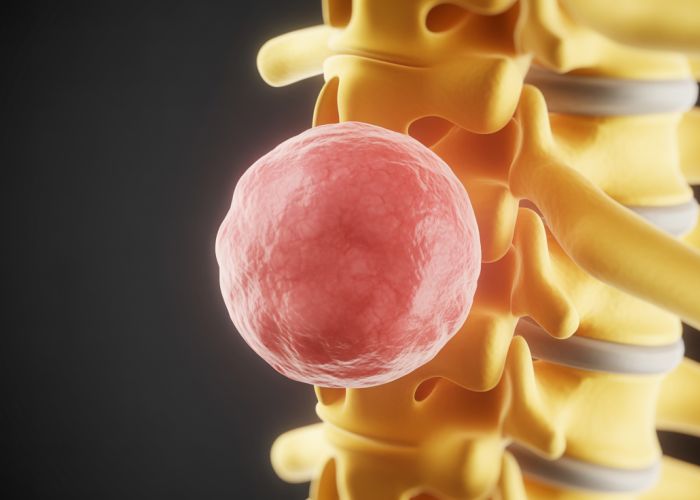
What exactly does a lipoma on the spine look like in pictures?
Lipoma on spine pictures often show a rounded or oval-shaped mass. Its appearance varies depending on its size and location. It may appear subtle, especially in early stages, or more prominent as it grows. Viewing lipoma on spine pictures provides a visual understanding of the potential size and shape.
How are lipoma on spine pictures used in diagnosis?
While lipoma on spine pictures from imaging techniques like MRI are crucial, they aren’t the sole basis for diagnosis. Doctors correlate images with physical examinations and patient symptoms. These lipoma on spine pictures help determine the size, location, and involvement of surrounding tissues.
What can lipoma on spine pictures reveal about potential treatment options?
The details visible in lipoma on spine pictures are essential for planning treatment. They show the tumor’s exact location and proximity to nerves or the spinal cord. This information helps surgeons determine if complete removal is possible and the best approach to minimize risks. Therefore, quality of lipoma on spine pictures is very important.
Do all lipoma on spine pictures show the same type of growth?
No, lipomas can vary. Some lipoma on spine pictures might show well-defined, encapsulated growths, while others might depict lipomas infiltrating surrounding tissues. The pictures also reveal if the lipoma is intradural (within the spinal cord covering) or extradural (outside the spinal cord covering), influencing management strategies.
So, hopefully, this gave you a better understanding of what lipoma on spine pictures are all about! If you’re concerned about anything you’ve seen or read, chatting with your doctor is always the best move. Take care!