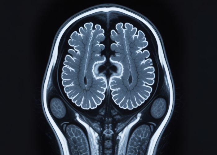
Cerebral Toxoplasmosis, an opportunistic infection of the brain, presents unique challenges in diagnosis, often requiring advanced imaging techniques. Magnetic Resonance Imaging (MRI), a cornerstone in neuroradiology, offers crucial insights into the characteristics of this infection. Understanding the nuances of mri picture cerebral toxoplasmosis necessitates a comprehensive knowledge of lesion morphology, a key attribute visualized in MRI scans. Immunocompromised individuals are particularly vulnerable to this condition, emphasizing the significance of early and accurate diagnosis. Leading medical centers rely on the detailed information provided by mri picture cerebral toxoplasmosis to formulate effective treatment plans and improve patient outcomes.

Understanding MRI Findings in Cerebral Toxoplasmosis
This article aims to explain how Magnetic Resonance Imaging (MRI) helps in diagnosing cerebral toxoplasmosis, focusing on what "mri picture cerebral toxoplasmosis" reveals.
What is Cerebral Toxoplasmosis?
Cerebral toxoplasmosis is a brain infection caused by the parasite Toxoplasma gondii. While many people are infected with this parasite, it rarely causes problems in individuals with healthy immune systems. However, in those with weakened immune defenses, such as individuals with HIV/AIDS or those undergoing immunosuppressive therapy, it can lead to serious illness affecting the brain.
The Role of MRI in Diagnosis
MRI is a crucial imaging technique for diagnosing cerebral toxoplasmosis. It provides detailed pictures of the brain, allowing doctors to identify characteristic lesions (abnormal areas) associated with the infection. While not definitive on its own, MRI in conjunction with clinical history and other diagnostic tests helps confirm the presence of the disease and guide treatment decisions.
Common MRI Findings in Cerebral Toxoplasmosis
Here are the common features doctors look for in an "mri picture cerebral toxoplasmosis":
Location of Lesions
- Basal Ganglia: One of the most frequent locations for lesions.
- Corticomedullary Junction: The area where the brain’s outer layer (cortex) meets the inner white matter.
- Thalamus: A central relay station for sensory and motor signals.
- Less Common Sites: Brainstem, cerebellum, and spinal cord.
Number and Size of Lesions
- Multiple Lesions: Cerebral toxoplasmosis often presents with multiple lesions distributed throughout the brain.
- Variable Size: Lesions can vary in size, from a few millimeters to several centimeters.
Appearance on Different MRI Sequences
MRI uses different "sequences" to highlight different tissue characteristics. The appearance of lesions varies depending on the sequence used. Understanding these differences is key to interpreting an "mri picture cerebral toxoplasmosis".
-
T1-weighted Images: Lesions may appear as areas of low signal intensity (darker than surrounding brain tissue), or occasionally isointense (similar signal intensity) before contrast administration.
- After Contrast (Gadolinium): Ring enhancement is a classic, but not always present, feature. This means the edges of the lesion become brighter after contrast is injected. The enhancement can be nodular or homogenous, but ring enhancement is highly suggestive when combined with location and clinical history.
-
T2-weighted Images: Lesions typically appear as areas of high signal intensity (brighter than surrounding brain tissue). This indicates edema (swelling) and inflammation around the lesions.
-
FLAIR (Fluid-Attenuated Inversion Recovery) Images: This sequence is sensitive to fluid and is particularly useful for detecting edema surrounding the lesions. A bright halo around the lesion is frequently observed.
-
Diffusion-Weighted Imaging (DWI): This sequence detects the movement of water molecules in the brain. Some lesions may show restricted diffusion (bright signal on DWI and low signal on ADC map), suggesting necrosis (tissue death) within the lesion.
The Significance of Ring Enhancement
Ring enhancement after contrast administration on T1-weighted images is a significant finding, but it’s not specific to cerebral toxoplasmosis. Other conditions, such as brain abscesses, tumors, and demyelinating diseases, can also cause ring enhancement. However, the combination of ring-enhancing lesions in the typical locations (basal ganglia, corticomedullary junction), along with clinical context (immunocompromised patient), strongly suggests cerebral toxoplasmosis.
Differential Diagnosis
It’s important to note that the MRI findings of cerebral toxoplasmosis can resemble other conditions. Therefore, a differential diagnosis is essential. Some conditions that can mimic cerebral toxoplasmosis on MRI include:
| Condition | Key Differentiating Features |
|---|---|
| Primary CNS Lymphoma | Lesions often located near ventricles; may have more homogenous enhancement; often positive EBV PCR in CSF. |
| Progressive Multifocal Leukoencephalopathy (PML) | Lesions typically in white matter, often confluent; usually no mass effect or enhancement; associated with JC virus. |
| Brain Abscess | More likely to have a single, well-encapsulated lesion with a thick, smooth capsule. |
Importance of Clinical Correlation
The "mri picture cerebral toxoplasmosis" is only one piece of the diagnostic puzzle. It’s crucial to correlate the MRI findings with the patient’s clinical history, symptoms, and other laboratory tests. Blood tests to detect Toxoplasma gondii antibodies and lumbar puncture to analyze cerebrospinal fluid (CSF) are often performed to confirm the diagnosis. Stereotactic brain biopsy may be needed if the diagnosis is uncertain.
MRI Cerebral Toxoplasmosis: Frequently Asked Questions
These FAQs answer common questions about how MRI pictures can help identify cerebral toxoplasmosis.
How does an MRI picture help diagnose cerebral toxoplasmosis?
MRI pictures can reveal characteristic lesions or abnormalities in the brain associated with cerebral toxoplasmosis. These lesions often appear as ring-enhancing masses on the mri picture. Detecting these patterns helps doctors narrow down the diagnosis.
What specific features on an MRI are indicative of cerebral toxoplasmosis?
Look for multiple, ring-enhancing lesions, particularly in the basal ganglia, corticomedullary junction, and thalamus. The "ring enhancement" refers to a bright ring seen around the lesion after contrast dye is injected during the mri picture process. This is a hallmark of cerebral toxoplasmosis.
If the MRI picture is negative, does that rule out cerebral toxoplasmosis?
Not necessarily. While an MRI is a valuable tool, early or mild cases might not show obvious lesions. If there is strong clinical suspicion, further testing, such as a brain biopsy, might be needed to confirm or rule out cerebral toxoplasmosis, even with a clear mri picture.
Can an MRI picture differentiate cerebral toxoplasmosis from other brain infections?
MRI findings can be suggestive, but other conditions like lymphoma or abscesses can present similar features. Therefore, the mri picture of cerebral toxoplasmosis must be interpreted in conjunction with the patient’s clinical history, blood tests, and other diagnostic information to reach an accurate diagnosis.
So, next time you hear about cerebral toxoplasmosis and someone mentions mri picture cerebral toxoplasmosis, you’ll have a better idea of what they’re talking about! Hope this was helpful!


